In Ophthalmology OSCE stations, images of various eye diseases are shown, and candidates are asked to identify the disease and describe its treatment. The following are examples of common cases that may be presented:
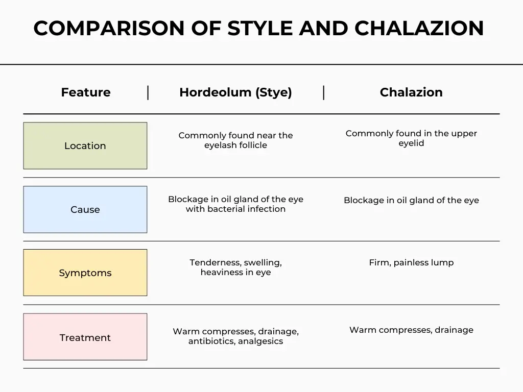
- Style (Hardeolum) and Chalazion
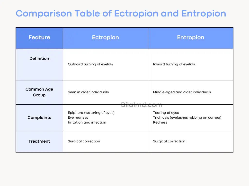
- Ectropion and Entropion
- Trachoma
- Structure of Eye:
- Conjunctivitis
- Dacrocystitis
- Endopthalmitis
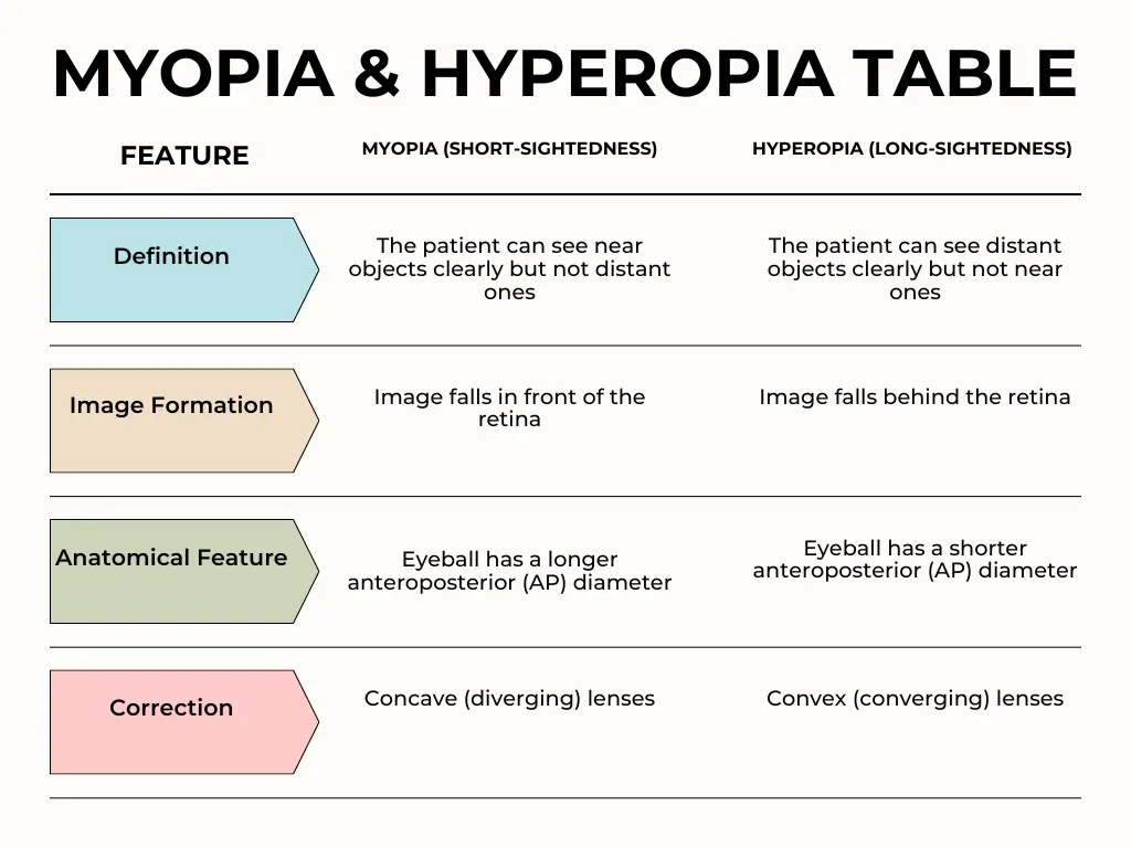
- Myopia and Hyperopia
- Cataract
- CRAO and CRVO
- Orbital Cellulitis
- Fundoscopic Finding in Diabetes Mellitus
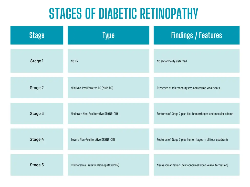
- STAGES OF DIABETIC RETINOPATHY
The Human eye consist of three layers.
- Outer Layer: Sclera and cornea
- Middle layer: Vascular in nature (choroid, iris)
- Nervous Layer: Retina (Tod and cones)
Segments of Eye:
- Anterial segment: Cornea to lens
- Posterior segment: Behind lens
- Anterior segment further divided into two chambers.
- Anterior chamber = cornea to iris
- Posterior chamber = iris to lens
1. Style (Hardeolum) and Chalazion
2. Ectropion and Entropion
3. Trachoma
A disease of eye caused by Bacterial infection which result visual impairment and even blindness.
Causes:
- Chlamydia Trachomatis
- It is contagious in nature
Symptoms:
- Mild itching and irritation in eye
- Photophobia
- Eyelid swelling
- Eye redness
- Vision loss
Who classification: (FISTO)
- TF: (Trachoma Follicular)
- Early infection heaving have more than five follicle.
- Follicle contain lymphocytes
- TI: (Trachoma Intense)
- In this stage eye are highly contagious and become irritated.
- TS (Trachoma Scarring)
- Repeated infection result scarring of inner eyelid.
- In this stage high chance of entropion
- TT (Trachoma Trichiasis)
- The scarred inner eyelid continue to deform and eventually turn inward (Entropion)
- TO: (Trachoma Opacity)
- In this stage clouding of cornea occur & result visual loss
- Tx: A single dose of Azihromycin.
4. Conjunctivitis
Inflammation of conjunctive in the eye. It is also know pink eye. It is usually due to infection or allergies
Types:
- Viral conjunctivitis (most common)
- Bacterial conjunctivitis
- Allergic conjunctivitis
Symptoms:
- Redness in eye
- Itchiness
- Gritty feeling
- Discharge (Purulent or watery)
- Photophobia
Treatment:
- Eye drop
- Eye ointment
- Anti-Allergic
- Antibiotics
5. Dacrocystitis
Inflammation of lacrimal sac present in nasolacrimal duct
Causes:
- Congenital
- Bacterial infection (Staph Aerous)
Symptoms:
- Swelling and near the medial canthus
- Tenderness
- Redness
- Pus discharge
- Fever
Diagnosis: Clinical diagnosis
Treatment:
- Oral antibiotic (7-10 days)
- DacryocystoRhinostomy (DCR)
6. Pterygium
- A non cancenus abnormal tissue growth on conjunctiva
- It usually grows toward cornea
Causes:
- Idiopathic
- Exposure to Uv-rays
- M>F
Diagnosis: Clinical diagnosed
Treatment:
- Follow up (cannot reach cornea)
- Surgery
7. Endopthalmitis
A purulent inflammation of intraocular fluid (Acquoud vs vitnus) due to infection usually after ocular surgery
Causes:
- Exogenous = Post-operative, Traumatic, Corneal Ulcer
- Endogenous = Bacterial, Fungal
Symptoms:
- Blurred Vision 94%
- Redness in eye 82%
- Pain in eye 74%
- Swollen lid
- Epiphora
O/E: Hypopyon
Note: Commonly occur after cataract surgery
Treatment:
- Corticosteriod
- Antibiotic
- Topical antibiotic
- Vitrectomy
8. Myopia and Hyperopia
9. Cataract
A condition in which the lens become opaque and cloudy. It is due to protein deposition during lens breakdown which result clouding.
Causes:
- Older Age
- D.M
- Trauma
- Ocular Injury
- Family History (e.g Congenital)
- Radiation
Types:
- Senile Cataract
- Nuclear Cataract
- Cortical cataract
- Posterior subcapsular cataract
- Congenital cataract
Treatment:
- Surgery ( IOL)
- Phacoemulsification
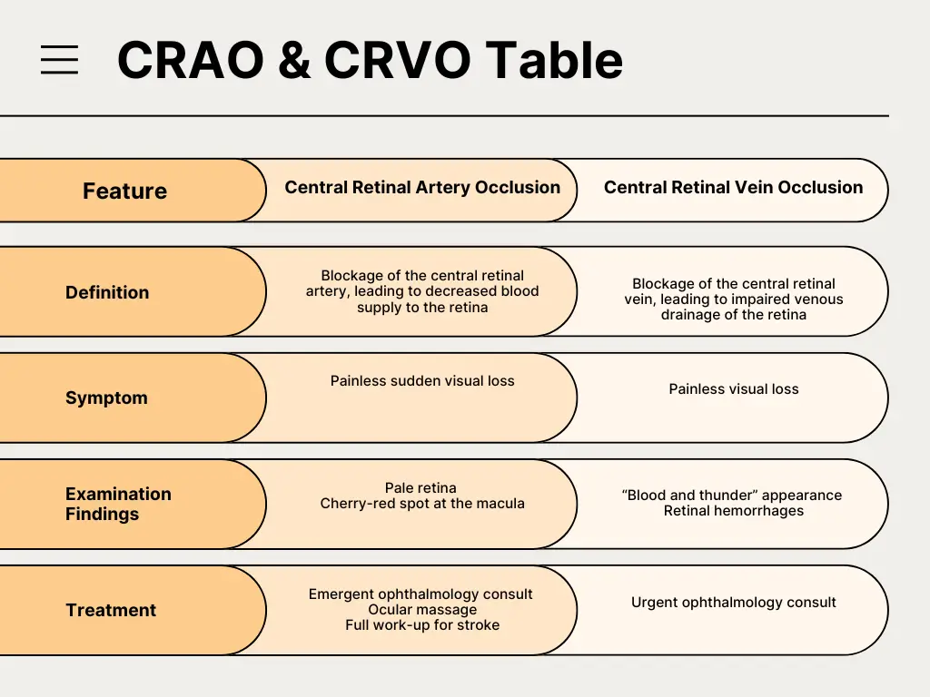
10. CRAO and CRVO
11. Orbital Cellulitis
Infection of fats and muscles around the eye. It affect the eye lids, eyebrows and cheeks
Cause:
Bacteria (Staph Aerous)
Symptoms:
- Proptosis
- Chemosis
- Pain with eye movement
- Fever
- Photophobia
Diagnosis:
Blood CP = Leukocytosis
Treatment:
Antibiotics, Corticosteroid
11. Fundoscopic Finding in Diabetes Mellitus
- Micro-aneurysm
- Cotton wool spot (Damage to nerve firbre)
- Hard exudate (yellow white deposits of protein & lipid)
- Retinal Hemorrhage
- Neovascularization