In the NLE Step-2 Dermatology exam, you won’t examine a live patient. Examiners typically present images either on a laptop or as printed photos, and you’ll have five minutes to complete each dermatology station. Be prepared for the high-yield cases that appear most frequently.
The following high-yield dermatology topics are asked repeatedly;
- Impetigo
- Contact Dermatitis
- Stasis Dermatitis
- Psoriasis
- Erythema Multiform
- Erythema Nodosum
- Molluscum Contagious
- Carbuncles
- Tinea Versicolor
- Tinea
- Basal Cell Carcinoma (BCC)
- Lipoma
- Seborrheic Dermatitis
- Acne Vulgarus
- Erysipelas
- Scabies
- Vertiligo
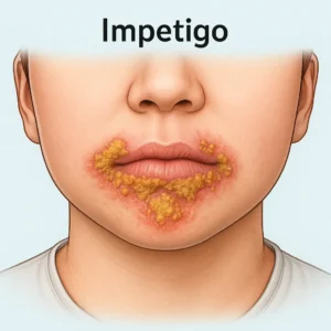
1. Impetigo
A superficial skin infection, which mainly involves the epidermis layer of skin. It is a highly contagious infection that affects infants & young children.
Causes:
- Streptococcus
- Staphylococcus aureus (Bullous)
Features:
A reddish sore around the nose and mouth which quickly ruptures, oozes for a few days, and then forms “Honey Coloured Crust”. A less common form is bullous impetigo.
Diagnosis:
- Clinical
Treatment:
- Mupirocin Ointment
2. Contact Dermatitis
A skin redness that results from contact with an allergen to which the patient has previously been exposed or sensitized.
The following are examples of contact dermatitis:
- Watch
- Rings
- Nickel
- Shoes
Diagnosis:
- Clinical diagnosis
- Patch test
Treatment:
- Topical corticosteroid
- Allergen avoidance
- Corticosteroid (severe cases)
3. Stasis Dermatitis
Skin changes occur as a result of stasis of blood due to impaired venous drainage.
Causes:
- Poor Circulation
- Value incompetency
- Venous insufficiency
Symptoms:
- Leg swelling
- Heaviness in the leg
- Pain that worsens with standing
- Skin ulcer
Treatments:
- Leg elevation
- Compression stocking
- Emollient
- Topical steroid
4. Psoriasis
A T-cell-mediated inflammatory condition, which is characterized by skin thickening and plaque.
Location:
Mainly affects the extensor part of the body (elbow + knee)
Lesion:
Typical lesion is “Demarcated Plaque with Silvery Scale”
Diagnosis:
- Clinical
Treatment:
- Local disease
- Topical steroid
- Vit-D
- Acitretin
- Methotrexate
- Anti-TNF Agents (Etanercept, Infliximab)
5. Erythema Multiform
A cutaneous reaction triggered by infection (HSV or mycoplasma)
Types of erythema multiform
There are two types of erythema multiform, as follows;
- Minor Form: Uncomplicated and localized to the skin.
- Major form: Involve mucous membrane, Systemic symptoms like fever, arthralgia, etc.
Treatment:
- Antiviral medications
- Antibiotics
Nikolsky Sign
Removing the epidermis through gentle rubbing.
Causes:
- Staphylococcal scalded skin syndrome (SSSS)
- Steven-Johnson Syndrome (SJS)
- Toxic epidermal Necrolysis (TEN)
- Pemphigus Vulgarus
6. Erythema Nodosum
Painful reddish nodules appear on the patient’s anterior shin. It is triggered by infection, a drug (OCP, sulfonamide, antibiotic)
and chronic inflammatory disease (TB and Sarcoidosis)
Treatment:
- Investigate and treat the underlying cause
- Cool compresses
- Bed Rest
- NSAID
7. Molluscum Contagiosum
It is a viral infection, mainly affecting children and immunocompromised patients.
Causes
- Caused by the poxvirus
Classic presentation:
- A tiny flesh coloured dome shaped lesion with central umbilication.
- Note: Typically, spare palm and sole.
8. Carbuncles
Infection of the hair follicle, which results in pus formation. Usually affects the back of the neck. Repeated attacks of carbuncles ruled out immune compromised state.
Treatment:
- Incision and Drainage
9. Tinea Versicolor
A condition caused by fungi that gives rise to different coloration.
Cause:
- Malassezia further
Presentation:
- Small patchy scaly lesion of varying colour.
Diagnosis:
- KoH preparation “Spaghetti and meatball appearance”
Treatment:
- Topical ketoconazole
10. Tinea: Known as Ringworm
Subtype:
- Tinea corporis
- Tinea Capitis
- Tinea Unguium
- Tinea Cruris
- Tinea Pedis
Lesion:
Erythematous border with central clearing.
Diagnosis:
- Clinical
- Best Initial-Potassium hydroxide to help identify fungal infections in skin, hair, or nail samples
- Most accurate = fungal
Treatment:
- Topical anti-fungal
- Oral griseofulvin
11. Basal Cell Carcinoma (BCC)
- A skin cancer which slow slow-growing, locally destructive, but has no metastatic potential.
- Most common skin cancer.
- The strongest risk factor is exposure to sunlight.
Types:
- Nudular
- Superficial
- Sclerosing
Diagnosis:
- Shave Biopsy
Treatment:
- Excision
- Cryotheraphy
- Mohs Surgery
12. Lipoma
- A round oval shaped lump of tissue that grows under the skin.
- It is made of fats and moves easily, and does not cause pain.
Symptoms:
The swelling is
- Painless
- Moveable
- Oval shape
- <2 inches in Diameter
Diagnosis:
- Clinical
- Biopsy to rule out cancer
- CT or MRI
Treatment:
- No Treatment required
- Surgically remove
13. Seborrheic Dermatitis
Fungal infection (Malassezia Spp)
Symptoms:
- Scaling and Crust
- Cradle Cap severe form, and it can bleed
Treatment:
Emmulin(vaseline)
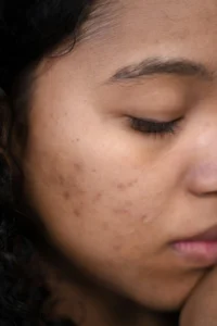
13. Acne Vulgarus
Its a common skin condition that occurs when hair follicles become clogged with oil, dead skin cells, and bacteria, It can cause a variety of symptoms, including: blackheads, whiteheads, pimple, and painful nodules under the skin.
14. Erysipelas
Superficial bacterial skin inv=fection that presents with well demarcated redness, swelling, and warmth.
Cause:
Group A streptococcus.
Treatment:
- Amoxicillin
- penicillin
15. Scabies
A contagious skin condition caused by sarcoptes scabiei (mite) tha burrows into the skin
Symptoms
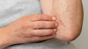
- Intense itching, especially at night
- Small, red papules or vesicles in typical sites
- interdigital spaces of fingers
- Wrists
- Genital area (in adults)
- Burrow marks
- Crusted scabies: Severe form in immunocompromised individuals
Diagnosis:
- Clinical presentation.
- Skin scraping showing mites, eggs, or fecal matter under the microscope
Treatment
- Permethrin 5% cream
- Oral treatment: Ivermectin
- Treat all close contacts simultaneously
- Wash clothing and bedding
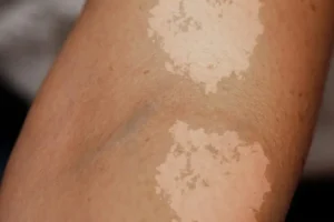
16 Vertiligo
- Autoimmune destruction of melanocytes
- White or depigmented patches on the skin