1. Spirometry (Pulmonary Function Test – PFT)
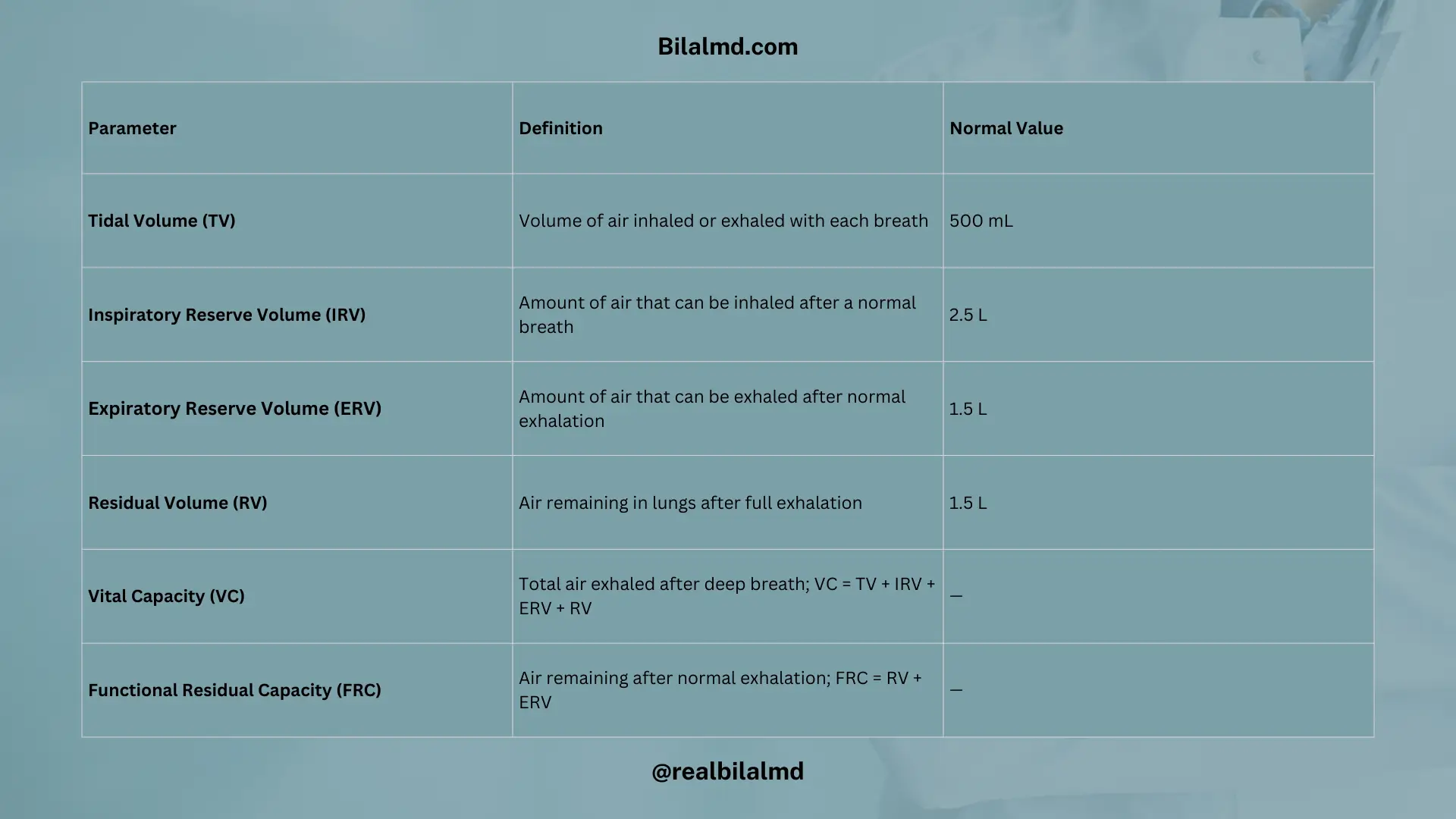
1. Tidal Volume (TV):
- Volume of air inhaled or exhaled with each breath.
- Normal Value: 500 mL.
2. Inspiratory Reserve Volume (IRV):
- The amount of air that can be inhaled after a normal breath.
- Normal Value: 2.5 L (Note: You mentioned 25 L, which seems to be a typo. Typical value is around 2-3 L).
3. Expiratory Reserve Volume (ERV):
- The amount of air that can be exhaled after a normal exhalation.
- Normal Value: 1.5 L.
4. Residual Volume (RV):
- The amount of air remaining in the lungs after a full exhalation.
- Normal Value: 1.5 L.
5. Lung Volumes and Capacities
1. Vital Capacity (VC):
- The total amount of air that can be exhaled after taking a deep breath.
- Formula: VC = TV + IRV + ERV + RV.
2. Functional Residual Capacity (FRC):
- The volume of air remaining in the lungs after a normal exhalation.
- Formula: FRC = RV + ERV.
2. Obstructive Lung Disease (ABCD)
- Asthma
- Bronchiectasis
- COPD (Chronic Obstructive Pulmonary Disease)
- Obstructive Lung Disease (general term)
These conditions typically involve increased airway resistance leading to difficulty exhaling, which is evident in reduced FEV1 (forced expiratory volume in 1 second) and FEV1/FVC ratio (forced vital capacity).
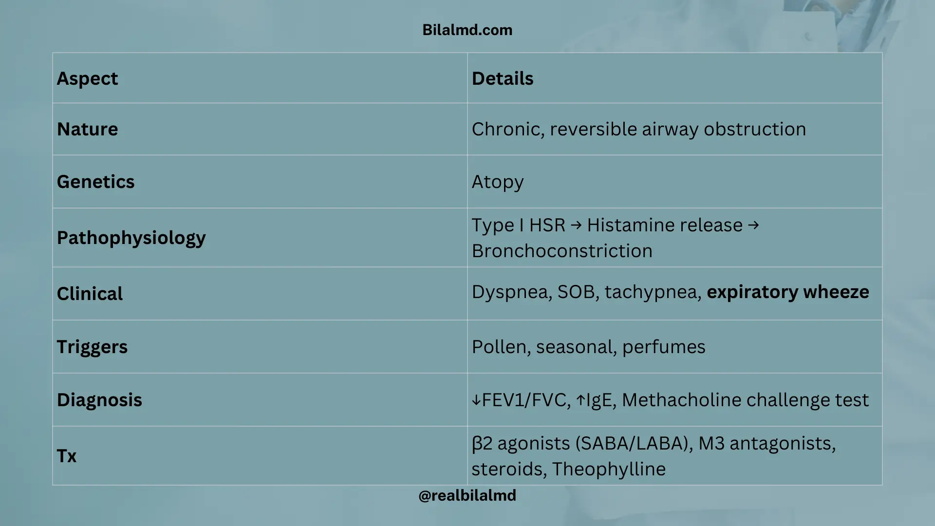
3. Asthma
Chronic, inflammatory, reversible airway obstruction.
1. Genetic Association:
- Atopy (genetic predisposition to develop allergic diseases such as asthma).
2. Pathophysiology:
- HSR-1 (Type I Hypersensitivity Reaction): Mediated by histamine, leading to inflammation and bronchoconstriction.
3. Clinical Presentation (P/C):
- Dyspnea: Difficulty breathing.
- Shortness of Breath (SOB): More common in childhood.
- Tachypnea: Increased respiratory rate, typically observed in adulthood.
- Expiratory Wheeze: Audible wheezing on exhalation, a hallmark of asthma.
- Can occur in both childhood and adulthood.
4. Triggers:
- Pollen.
- Seasonal changes.
- Perfume (allergic triggers).
5. Diagnostic Tests:
- FEV1/FVC ratio: Less than 70%, indicating obstructive lung disease.
- Methacholine challenge test: Positive in asthma, indicating airway hyperresponsiveness.
- IgE levels: Elevated, suggesting an allergic component to asthma.
6. Management of Asthma
- Bronchodilators:
- Beta-2 Agonists:
- SABA (Short-Acting Beta Agonists):
- Salbutamol, Albuterol.
- Action: Rapid relief of acute symptoms by relaxing bronchial muscles.
- LABA (Long-Acting Beta Agonists):
- Salmeterol, Formoterol.
- Side Effects: Tremor (a common side effect of beta-2 agonists).
- SABA (Short-Acting Beta Agonists):
- M3 Antagonists:
- Ipratropium, Tiotropium.
- Action: Reduce mucus production and help in bronchodilation.
- PDE Inhibitors:
- Theophylline.
- Side Effects: Cardiotoxic and neurotoxic effects, including arrhythmias and seizures, especially at high doses.
- Beta-2 Agonists:
4. Kartagener Syndrome (A type of Primary Ciliary Dyskinesia)
- Dextrocardia.
- Bronchiectasis.
- Sinusitis.
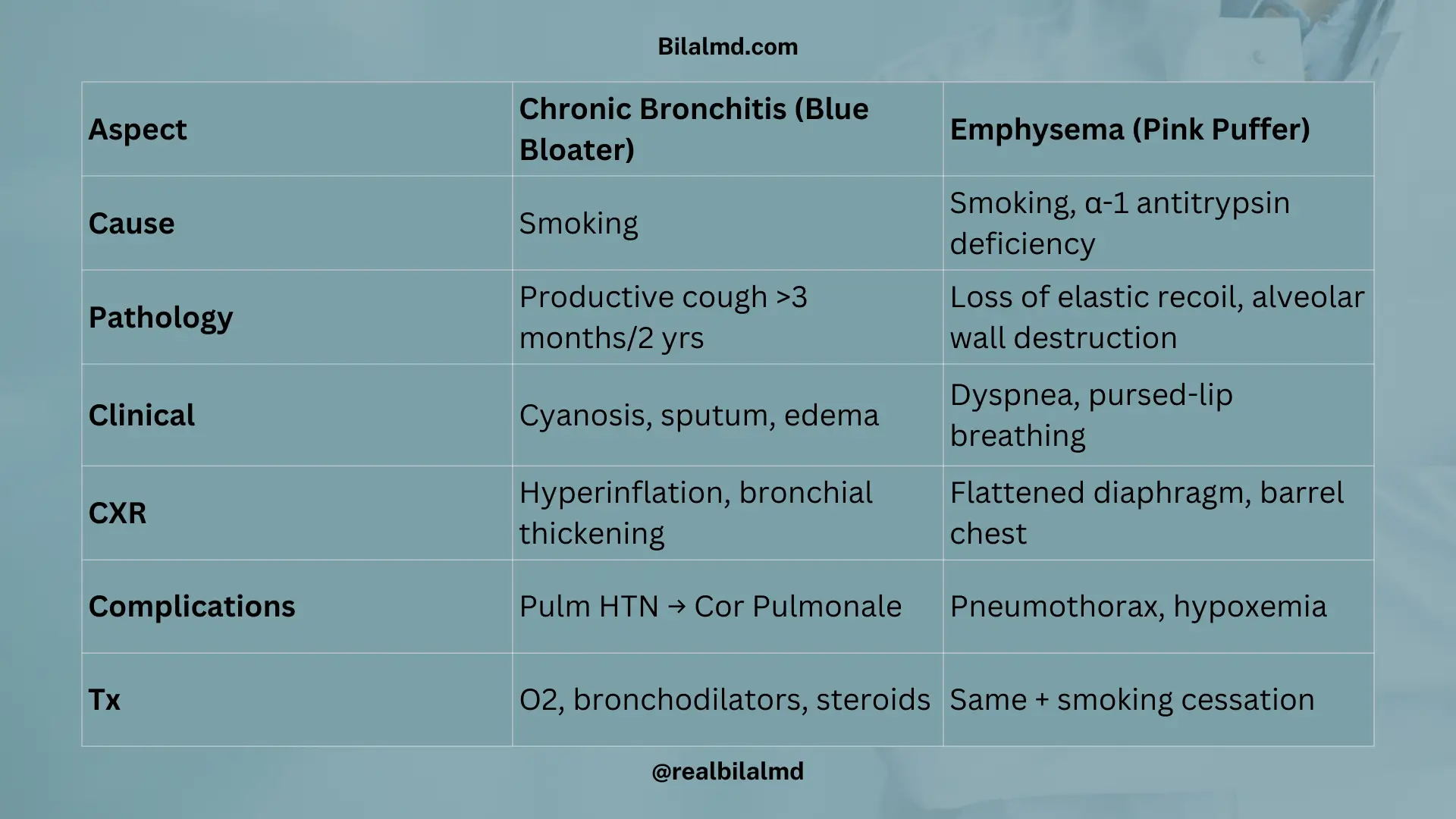
5. COPD (Chronic Obstructive Pulmonary Disease)
| Aspect | Chronic Bronchitis | Emphysema |
|---|---|---|
| Definition | Chronic, irreversible airway obstruction | Chronic, irreversible airway obstruction |
| Key Characteristic | Productive cough lasting >3 months for 2 consecutive years | Damage to alveolar septal walls, leading to loss of elastic recoil |
| Common Cause | Chronic smoking | Smoking (centriacinar), Alpha-1 antitrypsin deficiency (panacinar) |
| Chest X-ray | Abnormal changes (e.g., hyperinflation, bronchial wall thickening) | Hyperinflation and flattened diaphragm (Barrel-shaped chest) |
| Clinical Manifestations | Cough, sputum production, cyanosis | Shortness of breath, decreased breath sounds, pursed-lip breathing |
| Complications | Pulmonary hypertension → Right heart failure (Cor Pulmonale), Edema | Pneumothorax, right heart failure due to hypoxemia |
| Type of COPD | Blue bloaters (due to cyanosis and fluid retention) | Pink puffers (due to difficulty exhaling, using pursed lips) |
| Management | Oxygen therapy, bronchodilators, steroids | Bronchodilators, oxygen therapy, and smoking cessation |
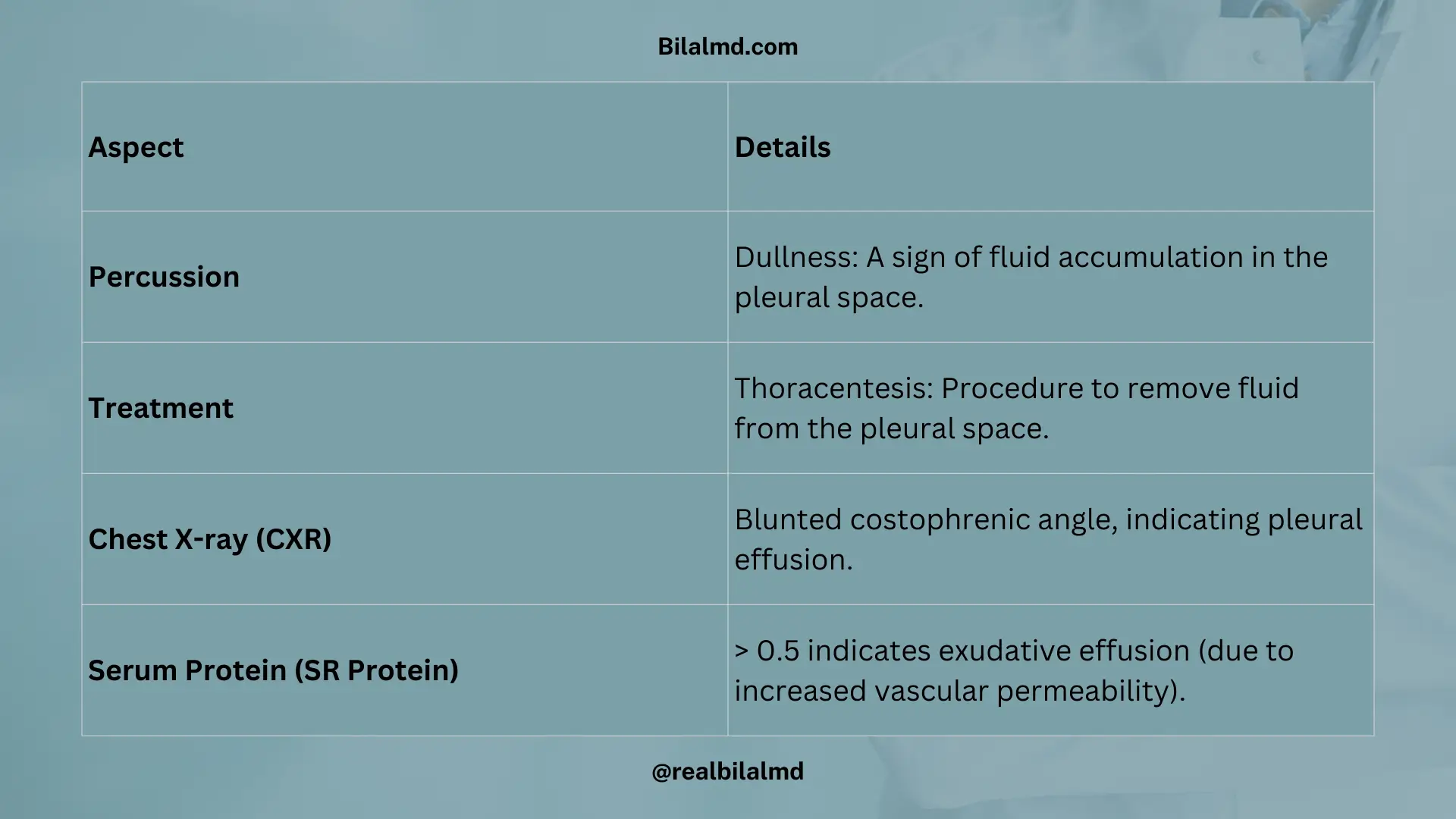
6. Pleural Pathology
| Aspect | Details |
|---|---|
| Percussion | Dullness: A sign of fluid accumulation in the pleural space. |
| Treatment | Thoracentesis: Procedure to remove fluid from the pleural space. |
| Chest X-ray (CXR) | Blunted costophrenic angle, indicating pleural effusion. |
| Serum Protein (SR Protein) | > 0.5 indicates exudative effusion (due to increased vascular permeability). |
7. Pneumothorax
| Aspect | Details |
|---|---|
| Definition | Pneumothorax occurs when air enters the pleural space and separates the lung parenchyma from the chest wall. |
| Tension Pneumothorax | Air accumulates only in the pleural space, causing tracheal deviation and hyper-resonance. This is a medical emergency. |
| Clinical Presentation | Tracheal shift (due to pressure buildup), hyper-resonance on percussion. |
| Recurrent Pneumothorax | Treated with chest tube insertion to evacuate air and prevent recurrence. |
8. Lung Cancer
- Leading Cause of Mortality:
Lung cancer is the leading cause of cancer-related mortality. - Risk Factors:
- Age: Common in individuals 50 years and older.
- Smoker: Strongly associated with a smoking history.
- Hemoptysis: Coughing up blood.
- Weight loss: Common symptom.
1. Ectopic Hormone Production in Lung Cancer:
- ACTH (Cushing Syndrome):
- Seen in small cell lung cancer (SCLC), leading to Cushing syndrome.
- ADH (SIADH):
- Seen in small cell lung cancer (SCLC), leading to Syndrome of Inappropriate Antidiuretic Hormone Secretion (SIADH).
- Cyclic Citrullinated Antibody (CCB):
- Associated with Lambert-Eaton Myasthenic Syndrome (LEMS) and Myasthenia Gravis (MG), often found in small cell lung cancer (SCLC).
2. Non-Small Cell Lung Cancer (NSCLC):
- PTH-rP (Parathyroid Hormone-related Protein):
- Causes hypercalcemia in squamous cell carcinoma of the lung.
- Typically occurs in non-smokers.
- Serotonin Increase:
- Seen in carcinoid tumors, which cause carcinoid syndrome (flushing, diarrhea, and wheezing).
Here are other materials for NLE NRE step 1
9. Sleep Apnea
Sleep apnea refers to the transient stoppage of breathing during sleep, which disrupts sleep quality.
Types of Sleep Apnea:
- Obstructive Sleep Apnea (OSA):
- Airway Obstruction: Blockage of the upper airway during sleep, leading to interrupted breathing.
- Causes:
- Adenoid and tonsillitis.
- Improper posture during sleep.
- Obesity.
- Central Sleep Apnea:
- Etiology: Specific causes related to the central nervous system (e.g., brainstem dysfunction).
9. Systemic Sarcoidosis
- Clinical Features:
- Erythema nodosum: Painful red nodules, typically on the shins.
- Hilar lymphadenopathy: Enlargement of lymph nodes in the chest.
- Diagnosis:
- Best Initial Test: Chest X-ray to assess for hilar lymphadenopathy.
- Confirmatory Test: CT-guided biopsy for tissue analysis.
- Diagnosis:
- Systemic Sarcoidosis.
- Treatment:
- Best Treatment: Corticosteroids (e.g., prednisone).
- Lofgren Syndrome:
- A form of sarcoidosis characterized by:
- Fever.
- Arthritis.
- Erythema nodosum.
- A form of sarcoidosis characterized by:
| Aspect | Silicosis | Asbestosis |
|---|---|---|
| Exposure | – Mine workers – Pottery workers – Silica exposure | – Tile workers – Building demolition – Shipbuilding |
| Key Features | – Eggshell calcification – Snowstorm appearance on imaging | – Calcified pleura – Plaque formation – Ground glass appearance |
| Risk | – Increases risk for Tuberculosis (TB) | – Increases risk for Bronchogenic carcinoma and Mesothelioma |
| Diagnosis | – Chest X-ray: Snowstorm appearance – CT scan: Eggshell calcification | – Chest X-ray: Pleural plaques – CT scan: Ground glass appearance |
| Common Diseases Associated | – Silicosis | – Bronchogenic carcinoma – Mesothelioma |
Check your NRE Step 1 result after completing the exam.