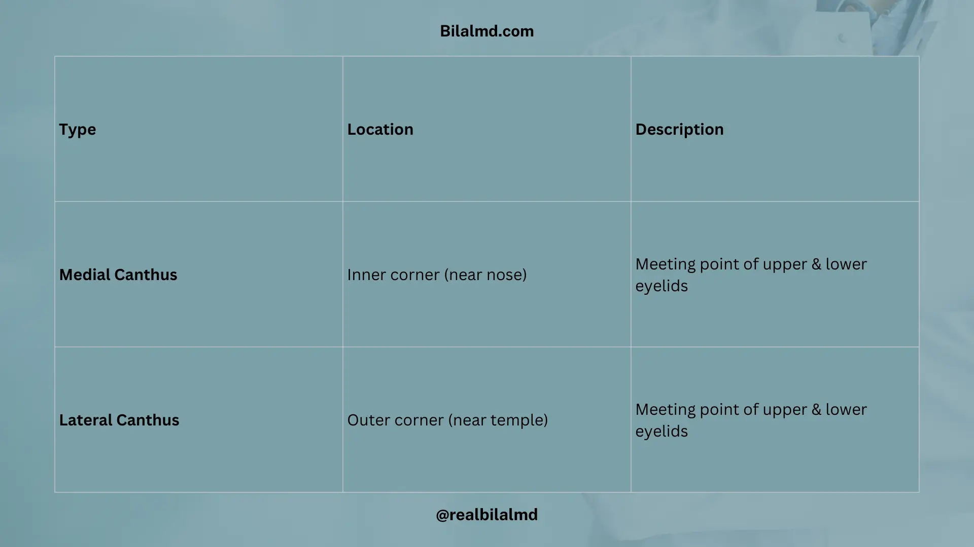
1. Canthus of the Eye
- Definition: The point where both eyelids meet.
- There are two canthi of the eye:
- Medial Canthus: The inner corner of the eye, where the upper and lower eyelids meet near the nose.
- Lateral Canthus: The outer corner of the eye, where the upper and lower eyelids meet near the temples.
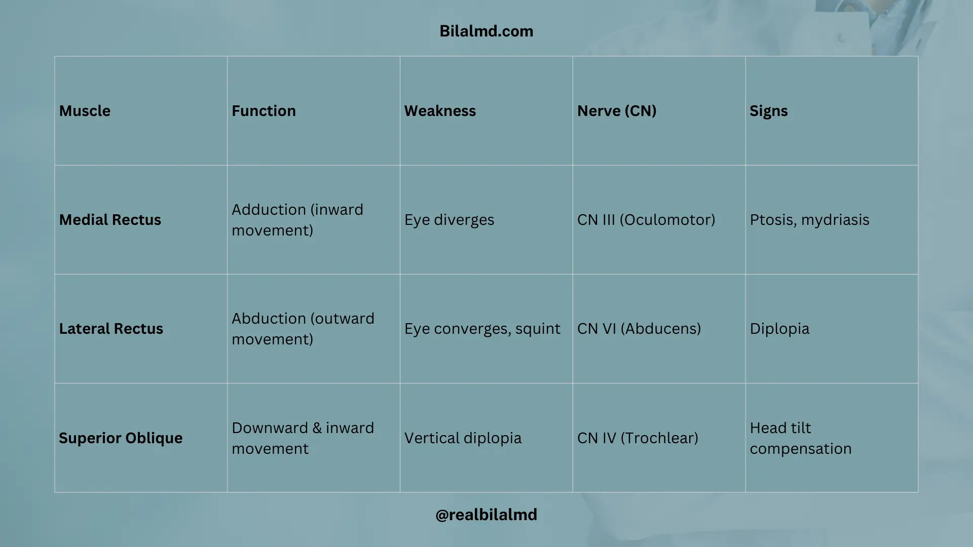
2. Eye Muscles and Nerve Control
- Medial Rectus:
- Function: Responsible for adducting the eye (moving it inward).
- Weakness: Causes the eye to diverge.
- Innervation: Controlled by the Oculomotor Nerve (CN III).
- Signs:
- Ptosis (drooping eyelid)
- Mydriasis (dilated pupil)
- Lateral Rectus:
- Function: Responsible for abducting the eye (moving it outward).
- Weakness: Causes the eye to converge and can lead to squint.
- Innervation: Controlled by the Abducens Nerve (CN VI).
Here are other materials for NLE NRE step 1
3. Superior Oblique Muscle and Down Syndrome
- Superior Oblique Muscle:
- Innervated by CN IV (Trochlear Nerve)
- Function: Helps with eye movement, allowing the eye to look downward and inward (important for focusing).
- Down Syndrome:
- The Superior Oblique Muscle is typically not affected in Down syndrome. The condition can impact other aspects of vision, but the specific nerve and muscle control for eye movements usually remain intact.
4. Eye Findings and Their Significance
- Bitot’s Spot:
- Location: Conjunctiva (white patches on the conjunctiva)
- Cause: Due to vitamin A deficiency, which is often seen in conditions like xerophthalmia.
- Roth’s Spot:
- Location: Retina
- Cause: Associated with infective endocarditis, these are retinal hemorrhages with a white center (often caused by emboli).

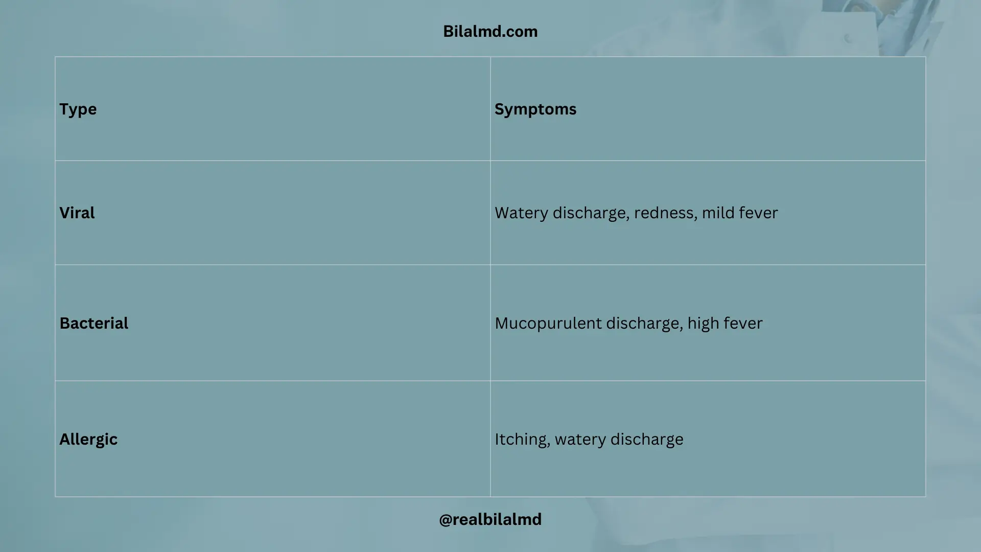
5. Conjunctivitis
1. Conjunctivitis Types and Symptoms
| Type | Symptoms |
|---|---|
| Viral | Watery discharge- Redness- Mild fever |
| Bacterial | Mucopurulent discharge (thick, yellowish)- Watery discharge- High fever |
| Allergic | Itching- Watery discharge |
2. Night Blindness
- Cause: Vitamin A deficiency
- Effect: Impaired ability to see in low light or darkness due to insufficient levels of retinal (a form of vitamin A) in the eye.
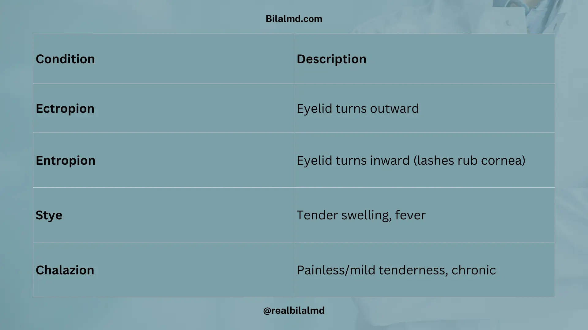
3. Eyelash and Eyelid Conditions
| Condition | Description |
|---|---|
| Outward (Ectropion) | Eyelid turns outward, causing the lower lid to fall away from the eye. |
| Inward (Entropion) | Eyelid turns inward, causing the lashes to rub against the cornea and eye. |
4. Eyelid Pathology
| Condition | Symptoms |
|---|---|
| Stye | Tenderness + Fever |
| Chalazion | Mild tenderness |
6. Glaucoma
Cause: Increased aqueous humor production or decreased drainage.
Types:
| Type | Description |
|---|---|
| Closed-Angle Glaucoma | More severe and faster progression Symptoms: blurred vision, redness, Pressure > 40 mmHg– Requires emergency care |
| Open-Angle Glaucoma | Iridocorneal angle does not drain properly- Drainage decreases, causing fluid retention and increased pressure |
Treatment:
| Medication | Action |
|---|---|
| Timolol | Beta-blocker, reduces aqueous humor production |
| Acetazolamide | Carbonic anhydrase inhibitor, reduces fluid production |
Surgical Treatment: Iridotomy: A surgical procedure to improve drainage.
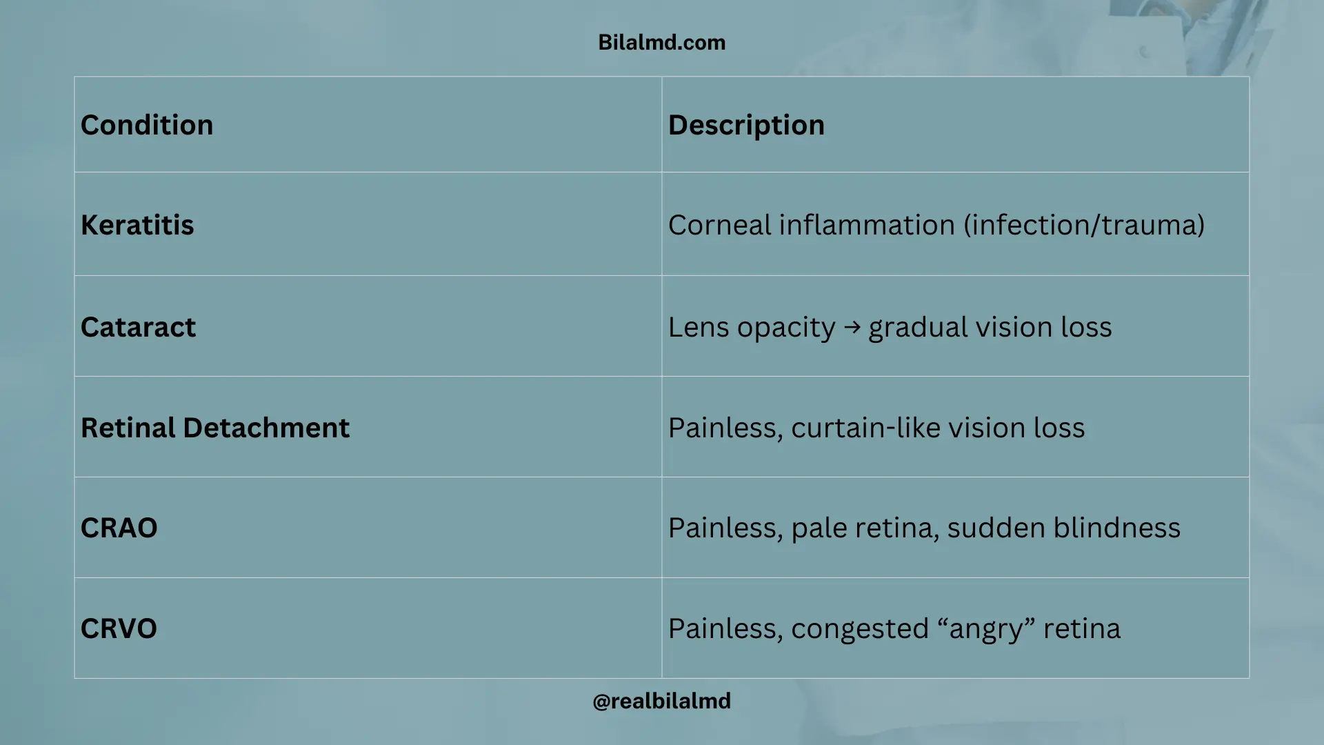
7. Cornea
| Condition | Description |
|---|---|
| Keratitis (Cornea) | Inflammation of the cornea, often caused by infection or injury. |
| Cataract (Lens) | Opacity in the lens, leading to decreased vision.- Senile Cataract: Most common in old age. |
| Retinal Detachment | Painless– Visual loss– Curtain-like shadow falling over vision. |
| Central Retinal Artery Occlusion | Painless– Visual loss– Pale retina due to lack of blood supply. |
| Central Retinal Vein Occlusion | Painless– Angry-looking retina due to blood congestion. |
1. Refractive Errors
| Condition | Description |
|---|---|
| Myopia | Near-sightedness– Can see near objects clearly but cannot see far objects. |
| Hyperopia | Far-sightedness– Can see far objects clearly but cannot see near objects. |
| Astigmatism | Blurred vision– Can also cause double vision due to irregular curvature of the cornea or lens. |
| Presbyopia | Age-related condition where the ability to focus on near objects decreases with age. |
Check your NRE Step 1 result after completing the exam.