Stroke volume calculator a fast, clinical way to get SV (mL/beat) and SVI. Enter what you have: EDV–ESV, CO ÷ HR, or echo’s LVOT area × VTI; the tool handles the math and can index to BSA (Mosteller) for size-adjusted insight. Use it when you need a trustworthy per-beat volume, not guesses from HR or BP alone.
Stroke Volume Calculator
Stroke Volume (SV): mL/beat
Cardiac Output: L/min
Cardiac Index (CI): L/min/m²
Stroke Volume Index (SVI): mL/m²/beat
Explore more heart calculator:
What is Stroke Volume?
Stroke volume (SV) is the amount of blood the left ventricle ejects with each beat (mL/beat). Conceptually, it’s one piece of cardiac output (CO), the other is heart rate (HR):
CO = SV × HR.
Typical resting adult SV is roughly ~60–100 mL (often taught around ~70 mL in a 70-kg male), but it varies with body size, age, loading conditions, and measurement technique.
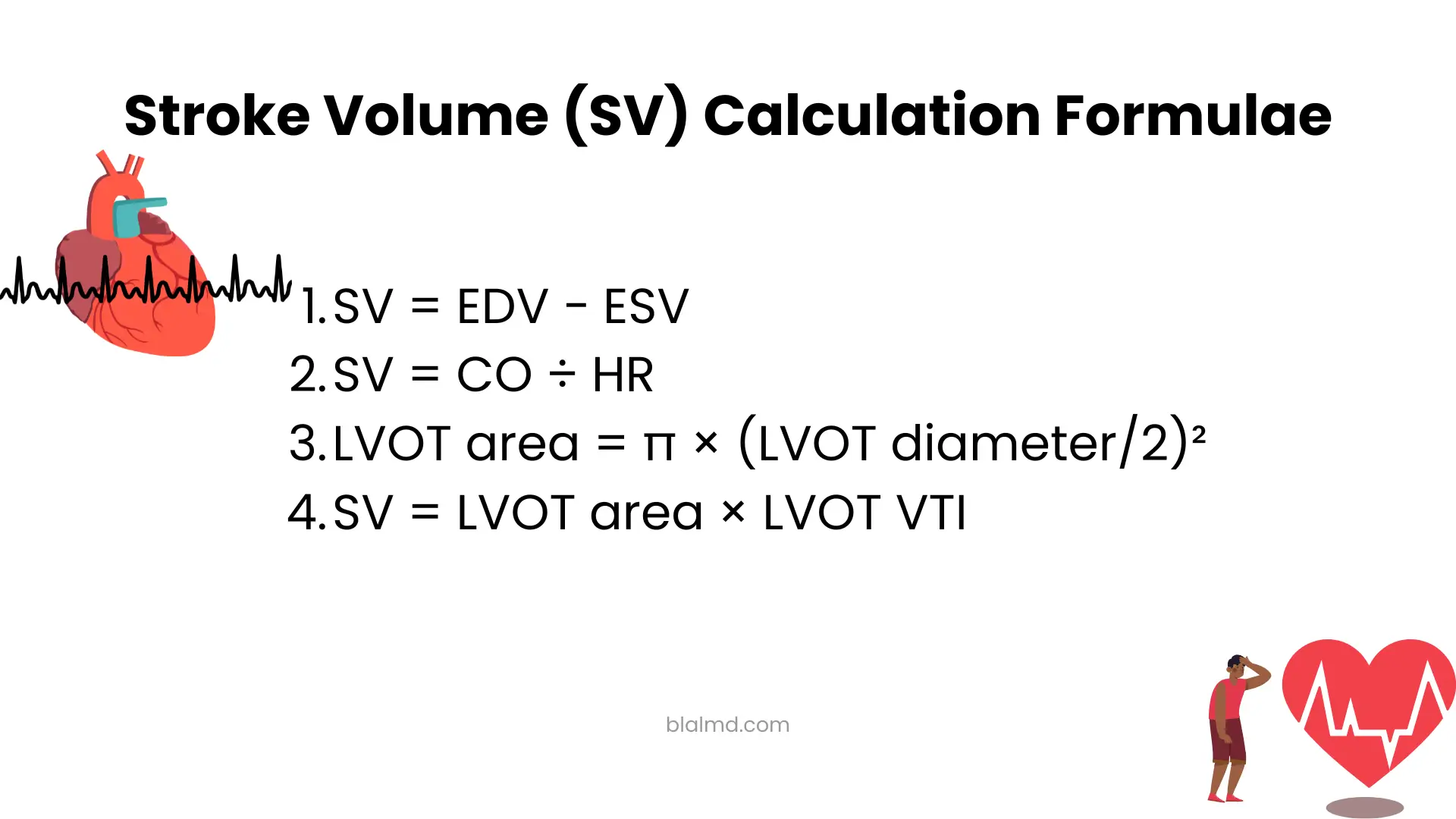
Stroke Volume Formula (All Valid Ways)
There isn’t just one “stroke volume formula.” Several are correct depending on the data you have:
- Volume method (ventriculography/echo/MRI):
SV = EDV − ESV (end-diastolic volume minus end-systolic volume). - From cardiac output and heart rate:
SV = CO ÷ HR. Use this when CO is measured (e.g., thermodilution, Fick, or echo) and HR is known. - Echocardiography (Doppler) using LVOT:
- LVOT area = π × (LVOT diameter/2)²
- SV = LVOT area × LVOT VTI (velocity–time integral)
This is the most common stroke volume echo calculation at the bedside.
These formulas are mathematically consistent. What changes is how you measured the inputs (volumes, CO, or Doppler signals).
How to Calculate Stroke Volume (Step-by-Step)
A) Echo (LVOT–VTI) example the practical “stroke volume echo calculator” approach
- Measure LVOT diameter in parasternal long-axis (cm).
- Compute LVOT area = π × (D/2)².
- Acquire LVOT VTI (cm) from apical 5-chamber or 3-chamber view.
- SV (mL) = LVOT area (cm²) × VTI (cm).
Worked example:
- LVOT diameter D = 2.0 cm → Area = π × (1.0)² ≈ 3.14 cm²
- VTI = 20 cm
- SV ≈ 3.14 × 20 = 62.8 mL
Round to 63 mL/beat.
Accuracy tips: LVOT is not perfectly circular; measurement errors in D are squared in the area calculation, so be meticulous. 2D assumptions and caliper placement matter.
B) CO/HR example when you already have cardiac output
If monitor or catheter says CO = 5.0 L/min and HR = 70 bpm:
- Convert CO to mL/min: 5000 mL/min
- SV = 5000 ÷ 70 ≈ 71 mL per beat.
C) EDV–ESV example when you have ventricular volumes
If EDV = 120 mL and ESV = 50 mL:
- SV = 120 − 50 = 70 mL.
Stroke Volume Index (SVI): Formula, Normal Values, and Why It Matters
SVI adjusts SV to body surface area (BSA) to allow fair comparison across body sizes:
SVI = SV ÷ BSA (mL/m²).
BSA is commonly computed using the Mosteller formula:
BSA (m²) = √(height(cm) × weight(kg) ÷ 3600).
Normal SVI commonly referenced around >35 mL/m²; values <35 mL/m² are used to flag low-flow states in contexts like aortic stenosis. (Ranges vary by population and technique.)
Example: Using the echo SV above (63 mL) and a 170 cm/70 kg adult:
- BSA = √(170×70 ÷ 3600) ≈ √3.306 ≈ 1.82 m²
- SVI = 63 ÷ 1.82 ≈ 34.6 mL/m² (borderline low depending on context).
“How to Calculate Stroke Volume from Heart Rate”
You cannot determine SV from HR alone. HR tells you beats per minute; you still need CO (then SV = CO/HR) or a method that directly measures flow/volume (e.g., echo LVOT–VTI). Any article implying HR alone is sufficient is misleading.
“How to Calculate Stroke Volume from Blood Pressure” What’s Possible vs. What’s Reliable
Textbook physiology relates pulse pressure (PP) to a volume change divided by arterial compliance (C):
PP ≈ SV / C → SV ≈ PP × C.
The catch: arterial compliance varies by age, disease, medications, and mean arterial pressure. That means you can’t get an accurate SV from brachial BP alone unless you know or assume C, which is often wrong. Even in research, using SV/PP as a compliance proxy is debated. Bottom line: at the bedside, don’t rely on BP-only SV formulas when accurate decisions matter use echo or a validated CO monitor.
Normal Ranges: Use Them Wisely
- SV (adult, technique-dependent): commonly taught around ~70 mL; healthy ranges are broad and vary with sex, size, and method.
- SVI: >35 mL/m² is often used as “normal-flow” in valve disease literature; <35 mL/m² suggests low flow (interpret in clinical context).
Remember: these are guides, not absolutes. Compare like with like same patient, same method, similar loading conditions.
How to Use the Stroke Volume Calculator
- Choose a method (you provide the inputs):
- Echo method: LVOT diameter + LVOT VTI.
- CO/HR method: Cardiac output + heart rate.
- EDV–ESV method: Ventricular volumes.
- Enter height and weight if you want SVI (the tool can compute BSA via Mosteller).
- The calculator outputs SV (mL/beat) and, if requested, SVI (mL/m²) and helps you interpret results versus typical reference values.
FAQs
What is the stroke volume formula?
Three valid forms: SV = EDV − ESV, SV = CO ÷ HR, or SV = LVOT area × VTI (echo). Use the one that matches your inputs.
How do I calculate stroke volume on echo (stroke volume echo calculation)?
Measure LVOT diameter (to get area) and LVOT VTI; multiply area × VTI. That’s the standard stroke volume echo calculator method.
How to calculate stroke volume from heart rate?
You can’t go from HR alone. If you have CO, then SV = CO ÷ HR. Otherwise, use echo or another validated flow method.
How to calculate stroke volume from blood pressure?
BP by itself isn’t enough. PP ≈ SV/C, and C (compliance) is unknown and variable so it’s not reliable for clinical use.
What is stroke volume index?
SVI = SV ÷ BSA (mL/m²). Many references use >35 mL/m² as normal flow (context dependent).
What is the stroke volume index formula for echo?
Compute SV by LVOT area × VTI, compute BSA (Mosteller), then SVI = SV/BSA.
What’s a normal stroke volume?
Often taught around ~70 mL (adult), but ranges vary with sex, size, and technique. Use SVI for size-adjusted interpretation.
“Stroke volume calculator” vs “stroke volume equation” what should I use?
Use the calculator when you want consistent math plus SVI and unit handling; use the equation when you already have clean measurements and just need a quick manual check.
Safety Note
Numbers aren’t diagnoses. If SV or SVI looks very low (and symptoms like hypotension, dyspnea, chest pain, confusion, cold, clammy skin are present), seek urgent clinical evaluation. Interpretation requires a full hemodynamic context and an understanding of the measurement method.
Sources
- StatPearls – Cardiac Output: definition and CO = SV × HR relation.
- StatPearls – Stroke Volume: definition, typical values.
- Echocardiography (LVOT–VTI) method: formulas and steps.
- LVOT geometry caveats (ellipticity/assumptions): echo measurement nuances.
- SVI thresholds in valvular disease literature: <35 mL/m² = low flow (context dependent).
- Mosteller BSA formula: original citation and clinical references.
- Pulse pressure & compliance (PP ≈ SV/C): physiological basis and limitations.