An MR Calculator helps clinicians quantify the severity of mitral regurgitation in a fast and consistent way. The most widely used approach behind a modern MR Calculator is the PISA method, which turns a few simple ultrasound measurements into three core numbers that matter at the bedside. Those numbers are the volume flow rate through the leaking valve, the effective regurgitant orifice area, and the regurgitant volume per heartbeat. Understanding what these values mean and how to measure them correctly lets you turn a raw color Doppler image into a clear, actionable assessment of mitral regurgitation.
MR Calculator
Inputs
Severity (combined)
Results
Volume Flow Rate (VFR)
Effective Regurgitant Orifice Area (EROA)
Regurgitant Volume (RVol)
Notes
Method & Limitations
Hemispheric PISA: VFR = 2πr²Vₐ; EROA = VFR/Vmax; RVol = EROA × VTI. No angle correction is applied.
Educational use only; not a substitute for clinical judgment.
Explore more heart calculator:
What an MR Calculator does
An MR Calculator based on the PISA method computes three quantities from four ultrasound inputs. The inputs are the PISA radius, the color Doppler aliasing velocity, the peak regurgitant velocity from continuous wave Doppler, and the velocity time integral of the regurgitant jet. The calculator then applies standard formulas:
- Volume flow rate equals two times pi times the square of the PISA radius times the aliasing velocity
- Effective regurgitant orifice area equals volume flow rate divided by peak jet velocity
- Regurgitant volume equals effective orifice area multiplied by the regurgitant jet velocity time integral
These relationships come directly from fluid continuity and are the backbone of many trusted clinical tools.
When to use an MR Calculator
Use an MR Calculator when you need a quantitative measure of mitral regurgitation rather than a purely qualitative impression. PISA derived measurements are most helpful in primary mitral regurgitation caused by leaflet disease, in routine follow up of established mitral regurgitation, and in cases where treatment decisions depend on objective grading. Guidelines recommend an integrated approach that combines multiple signs and measurements. A calculator gives you the quantitative core of that approach and reduces subjective variation from one examination to the next.
Inputs you will need
PISA radius r
Measure from the center of the regurgitant orifice to the first aliasing line on the color Doppler hemisphere. If the hemisphere is too small, slightly lower the Nyquist limit to enlarge it. Most labs set aliasing velocity around thirty to forty centimeters per second to make the hemisphere easier to see and measure.
Aliasing velocity Va
This is the color Doppler Nyquist limit at which color reversal occurs. Record the value in centimeters per second and pair it with the radius measurement taken at the same time point in the cardiac cycle.
Peak jet velocity Vmax
Obtain this from continuous wave Doppler tracing of the mitral regurgitant jet. Use the true peak value in centimeters per second.
VTI of the regurgitant jet
Trace the jet envelope to obtain the velocity time integral in centimeters. This integrates the velocity over systole and turns an area into a volume when multiplied by the effective orifice area.
How to calculate step by step
- Confirm units before you start. Work in centimeters and centimeters per second to keep the math clean.
- Compute the volume flow rate: VFR equals two times pi times r squared times Va. In these units, the result is cubic centimeters per second, which equals milliliters per second.
- Compute the effective regurgitant orifice area: EROA equals VFR divided by Vmax. The result is square centimeters.
- Compute the regurgitant volume per beat: RVol equals EROA times VTI. The result is milliliters per beat.
These steps are what an MR Calculator automates. They also explain why each input must be measured at the same phase of the cardiac cycle. If you measure radius at one moment and velocities at another, you introduce error and may misclassify severity.
A quick worked example
Imagine a radius of 0.9 cm, aliasing velocity of 35 cm per second, peak jet velocity of 500 cm per second, and VTI of 120 cm.
- VFR equals 2 × 3.1416 × 0.9 × 0.9 × 35 which is about 177 mL per second
- EROA equals 177 divided by 500 which is about 0.35 cm²
- RVol equals 0.35 times 120 which is about 42 mL per beat
This example lands near the boundary between moderate and more severe disease and shows how small changes in measurements can shift categories. The goal of an MR Calculator is to anchor these decisions with transparent numbers.
Interpreting the results
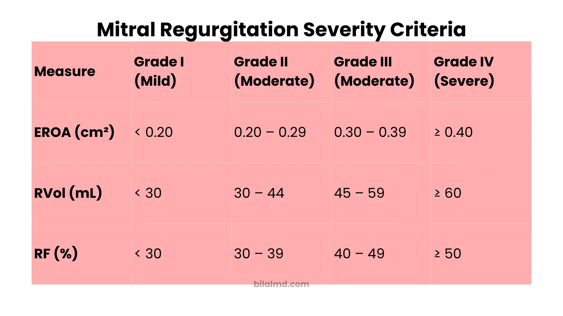
Two numbers usually drive clinical grading with a PISA based MR Calculator. These are EROA and regurgitant volume. Authoritative resources and society guidance documents use similar cutoffs for native primary mitral regurgitation. Severe disease is usually EROA at or above 0.40 cm² or regurgitant volume at or above 60 mL per beat. Values clearly below these levels support mild disease and values in between suggest a moderate range where clinical context matters.
Secondary or functional mitral regurgitation behaves differently because the orifice can be elongated and dynamic. European guidance updated the definition of severe secondary mitral regurgitation to an EROA of 40 mm² or greater which equals 0.40 cm², aligning it with thresholds used for primary disease. This change reflects outcome data and reduces past confusion about lower EROA thresholds for secondary cases. Always consider mechanism and geometry when you interpret PISA values in functional disease.
Guidelines also stress that you should not rely on one measurement alone. Combine quantitative outputs from an MR Calculator with other signs such as vena contracta width, pulmonary vein flow, mitral inflow patterns, and chamber response. This integrated method improves accuracy and is the standard way to grade severity in daily practice.
Practical tips that improve accuracy
Optimize the aliasing setting
Set the Nyquist limit to around thirty to forty centimeters per second to make the PISA hemisphere large and sharp. Then zoom in and freeze the frame that shows the hemisphere best for measurement.
Measure at the same moment in the cardiac cycle
Radius, aliasing velocity, and peak jet velocity should represent the same systolic moment. If you average across beats, do so consistently and note rhythm status.
Watch out for non hemispheric geometry
The PISA method assumes a hemispheric flow convergence. Eccentric jets, multiple jets, and orifices with significant ellipses or constraint may break that assumption. In these cases, your MR Calculator still produces a number but you should weigh it with caution and look for complementary measures.
Understand post procedure limitations
After edge to edge repair the orifice shape changes and PISA derived values have limited usefulness. Quantitative pulsed wave Doppler and three dimensional approaches can complement the assessment in that setting.
Complements and alternatives to PISA
Vena contracta width is a simple and fast metric. A small width suggests mild disease and a width near or above seven millimeters supports severe disease in primary cases. Biplane measurements and three dimensional vena contracta area can improve reliability when the orifice is not circular.
Volumetric methods that compare forward and total stroke volumes provide another way to compute regurgitant volume and regurgitant fraction. Emerging work suggests two dimensional volumetric echocardiography can outperform PISA in some primary disease scenarios. The choice of method depends on image quality, mechanism, and operator experience. An MR Calculator based on PISA remains valuable, but do not ignore a better suited method when your images support it.
Who benefits from an MR Calculator
Clinicians in echocardiography labs use an MR Calculator to produce reproducible numbers for reports and to guide decisions about timing of follow up or referral for surgical or transcatheter therapy. Trainees use it to learn the relationships between color Doppler images and valve physics. Patients benefit because objective numbers reduce variation and help explain why a plan is changing. Many professional resources point clinicians toward quantitative grading whenever possible rather than relying on descriptive language alone.
Step by step workflow for everyday use
- Acquire high quality color Doppler images in the best view for the jet
- Adjust aliasing to grow a clear hemisphere and measure the radius precisely
- Capture continuous wave Doppler of the same jet and trace the envelope for peak velocity and VTI
- Enter radius, aliasing velocity, peak velocity, and VTI into the MR Calculator
- Record VFR, EROA, and regurgitant volume
- Interpret results together with vena contracta width, pulmonary vein flow, and chamber response
- Document rhythm, heart rate, blood pressure, and any technical limitations that might influence the numbers
Following a consistent workflow like this one improves repeatability from study to study and between readers.
Frequently asked questions
Is a PISA based MR Calculator accurate enough for decisions about surgery or transcatheter repair
A well performed PISA calculation is part of the standard toolkit and is recommended as one component of an integrated assessment. It should be considered alongside other measurements and the clinical picture. When image quality or geometry is poor, complement it with volumetric or three dimensional measurements.
What settings should I use for the Nyquist limit when measuring the PISA radius
Many labs use an aliasing velocity near thirty to forty centimeters per second to create a larger and cleaner hemisphere. Adjust in small steps to avoid losing definition.
Are the cutoffs the same for primary and secondary mitral regurgitation
Today many authorities align severe secondary mitral regurgitation with an effective orifice area of at least 0.40 square centimeters, the same as primary disease. Past thresholds were lower for functional disease which caused confusion, so verify which guidance your team follows.
Which number is more reliable, EROA or regurgitant volume
Neither should stand alone. EROA and regurgitant volume offer different views of severity. Use both when available and integrate with other imaging signs as recommended in major guidelines.
How does an MR Calculator compare with volumetric methods
PISA based calculators are fast and widely available. Volumetric methods can be more accurate in selected cases of primary disease when images are ideal. The best approach is to use the method that fits the anatomy and the data quality in front of you.
Can I use an MR Calculator in atrial fibrillation
Yes, but average multiple beats and try to align measurements to the same part of the cycle. Irregular rhythm increases variability, so an integrated approach is even more important.