If you’re here, you’re not looking for fluff; you want a clear, medically grounded explainer that supports you and helps interpret their results. Here it is.
Cardiac Index Calculator
Yes No
Disclaimer: This cardiac index calculator provides estimates based on the input data. The results are intended for informational purposes only and should not be used as a substitute for professional medical advice. Always consult a healthcare provider for diagnosis and treatment.
Sources: NCBI – Cardiac Index, NCBI – Haycock Formula.
Explore more heart calculator:
What is cardiac index (CI)?
Cardiac index (CI) is cardiac output (the volume of blood your heart pumps each minute) normalized to body surface area (BSA), allowing for comparison of perfusion relative to body size. In plain terms: CI = CO ÷ BSA, reported in L/min/m². Normal CI at rest is typically ~2.5–4.0 L/min/m². Values under ~2.2 L/min/m² in a hemodynamically supported patient strongly suggest cardiogenic shock an emergency.
Why index instead of just “cardiac output”? Two people can have the same CO, but different body sizes; the smaller person may actually be better perfused. CI corrects that.
The cardiac index formula
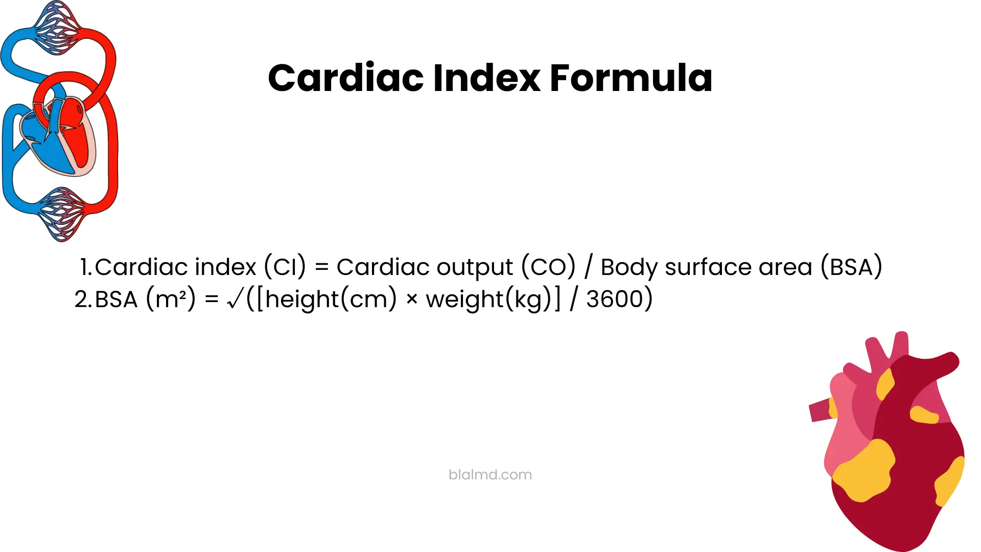
- Cardiac index (CI) = Cardiac output (CO) / Body surface area (BSA).
- CO itself comes from heart rate × stroke volume or is measured directly with clinical methods (Fick, thermodilution, Doppler echo, etc.).
- The most common quick BSA method is the Mosteller formula:
BSA (m²) = √([height(cm) × weight(kg)] / 3600).
How to calculate cardiac index (step-by-step)
You need either:
- Cardiac output , and
- Height and weight to compute BSA.
Example:
- Height = 170 cm, Weight = 70 kg → BSA = √(170×70/3600) = √(11900/3600) ≈ √3.3056 ≈ 1.818 m² (Mosteller).
- Suppose CO = 5.0 L/min (typical resting output).
- CI = 5.0 ÷ 1.818 ≈ 2.75 L/min/m², which sits in the normal range (2.5–4.0).
In practice, CO can be measured using the Fick principle, thermodilution (pulmonary artery catheter), Doppler echocardiography, or less-invasive arterial waveform analysis; each method has its pros and cons, as well as context-specific accuracy.
Interpreting CI: what “low” or “high” actually means
- Normal CI: about 2.5–4.0 L/min/m² at rest.
- Low CI: <~2.2 L/min/m² (with support) is consistent with cardiogenic shock physiology and poor organ perfusion, think acute myocardial infarction with pump failure, severe myocarditis, decompensated heart failure, etc. This is ICU-level care.
- High CI: >~4.0 L/min/m² at rest points toward high-output states (e.g., severe anemia, thyrotoxicosis, AV fistula, liver disease, sepsis’ hyperdynamic phase, obesity-related vasodilation). “High” doesn’t mean “healthy” it often signals underlying pathology with low systemic vascular resistance.
When and why clinicians use CI
- Shock differentiation: CI helps separate cardiogenic/obstructive/hypovolemic (usually low CI) from distributive (often high or normal CI with low SVR) critical for choosing fluids, vasopressors, or inotropes.
- Heart failure management: CI trends guide escalation (diuretics, inotropes, mechanical support).
- Peri-operative and critical care: Therapy titration hinges on whether perfusion fails from low flow (CI) or low resistance.
Using the Cardiac Index Calculator
- Enter height and weight → the calculator computes BSA via Mosteller.
- Enter cardiac output if you have a measured value (from clinical monitoring).
- The tool returns CI in L/min/m² and places it relative to the normal 2.5–4.0 range so users can quickly see if a result is typical, low, or high.
Reality check: CI is not a self-diagnosis. Numbers without clinical context are just numbers. Interpretation belongs to clinicians who know the method used to measure CO, the patient’s preload/afterload/contractility, and the full hemodynamic picture.
Accuracy caveats (don’t ignore these)
- There is no single gold standard for CO/CI in every setting. Thermodilution, Fick, Doppler, MRI, and arterial waveform methods each have biases and situational limitations.
- Some devices lose accuracy at very low CI (<2.2) or in vasoplegic states (e.g., advanced liver disease, septic shock). Know your device’s blind spots.
- BSA formulas differ (Mosteller, Du Bois, Haycock, etc.). Mosteller is widely used because it’s simple and aligns well with more complex models for adults, but recognize that different BSA formulas will shift CI slightly.
Frequently Asked Questions (FAQs)
What is a “good” cardiac index?
For most resting adults, 2.5–4.0 L/min/m² is considered normal. Below that suggests inadequate flow relative to body size; above that may reflect high-output physiology. Context still matters.
How do I calculate cardiac index without invasive lines?
Use non-invasive CO (e.g., echocardiography/Doppler) and compute BSA from height/weight with Mosteller’s formula; then CI = CO ÷ BSA. Validate results clinically.
CI vs. CO what’s the difference?
CO is absolute flow (L/min). CI divides CO by BSA, letting you compare perfusion across different body sizes more clinically comparable.
What conditions lower CI?
Cardiogenic shock (e.g., large MI), severe valvular disease, advanced heart failure, profound hypovolemia, and obstructive causes (PE, tamponade) classically drive CI down.
What conditions raise CI?
Thyrotoxicosis, sepsis (hyperdynamic phase), significant anemia, AV fistulas/shunts, cirrhosis, and sometimes obesity-related vasodilation. High CI can still coexist with heart failure symptoms don’t assume “high = healthy.”
What’s the fastest BSA equation to use?
Mosteller: √(height(cm) × weight(kg)/3600). It’s widely adopted and simple.
What’s the threshold for cardiogenic shock?
In many references, CI < 2.2 L/min/m² (with pharmacologic or mechanical support) strongly aligns with cardiogenic shock physiology. Treat it as urgent.
Can lifestyle changes “fix” a low CI?
If CI is low because of volume depletion, fluids may help; if due to pump failure, patients may need inotropes or mechanical support decisions made by clinicians. Lifestyle matters long-term, but acute low CI is a medical problem first.
How accurate is the Fick or thermodilution method?
Both are well-established; thermodilution is common in the ICU, while Fick is conceptually straightforward but invasive in its classic form. Each has error sources (e.g., shunts, tricuspid regurgitation, oxygen consumption estimates).
Does a high CI always mean good perfusion?
No. In high-output heart failure, CI can be >4.0, yet tissues may still be under-perfused because systemic vascular resistance is too low.
Safety note
This calculator does not diagnose any condition. If your CI looks low (or very high) and you have symptoms (chest pain, breathlessness, dizziness, confusion, cold, clammy skin), seek urgent medical care. Numbers without context can mislead
References
- StatPearls: Physiology, Cardiac Index (formula, normal range, measurement methods, shock patterns).
- Mosteller BSA formula references and clinical calculators.
- High-output heart failure thresholds and causes (CI >4.0).
- Cardiac output measurement methods & considerations (Fick, thermodilution).
- Cardiogenic shock background and outcomes.