Pregnancy ultrasounds offer an exciting window into the womb, allowing parents and providers to watch a baby’s growth week by week. From the earliest confirmation of pregnancy to detailed anatomy scans and late-third-trimester checkups, each ultrasound reveals new milestones. We’ll also highlight how our Conception Calculator tool can use ultrasound findings to estimate gestational age and due dates – helping answer the common question, “How far along am I?”
First Trimester (Weeks 1–12)
The first trimester begins from the 1st day of the last menstrual period, from weeks 1 to 12 of pregnancy. Early on, the embryo is microscopic, so the initial weeks show little on ultrasound. By about week 5, an ultrasound can confirm the pregnancy’s location and viability. Through weeks 6–12, rapid development occurs with a heartbeat to a recognizably formed fetus by the end of the trimester. First-trimester ultrasounds are crucial to see vital organs in the pregnancy (measuring the embryo’s crown-rump length, CRL) and for early screenings.
Weeks 1–2 (Preconception and Ovulation)
In “week 1” of pregnancy, you’re technically not pregnant yet – this is the week of your last menstrual period. By week 2, ovulation occurs, and conception is possible. Ultrasound at 1–2 weeks pregnant does not show any embryo, as implantation hasn’t happened. The uterine lining may be thickening in preparation, but no gestational sac is visible this early. It’s normal at this stage that there’s nothing to see on an ultrasound. If implantation occurs menstrual period also stops.
Weeks 3 Fertilization
Sperms meet the egg that was released during ovulation. Both of them unite and form a new type of cell called a zygote that is then transported from the fallopian tube to the uterus for implantation. Before implantation zygote starts to divide and form a cluster of cells called a morula, then become blastocyte. An ultrasound in week 3 still won’t detect the embryo.
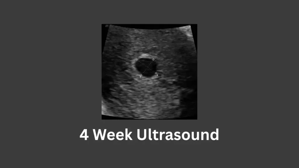
Weeks 4 Implantation
Now blastocyte start to attach to the uterus wall. We call this implantation. The inner cells of the blastocyte has zygote (baby) and the outer cells will become the placenta that will nourish the baby. A tiny black area in the uterus – the gestational sac – might be seen on a transvaginal ultrasound. This small fluid-filled sac is the first visible sign of pregnancy. The gestational sac measures only a few millimeters and may be barely detectable. The embryo itself is still too small to see.
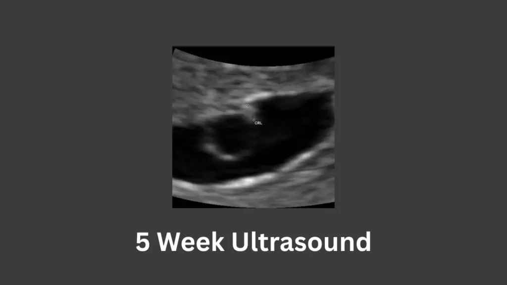
5 Weeks Hormone levels rise
Week 5 is often the earliest a pregnancy is confirmed by ultrasound. At a 5-week ultrasound, you will likely see the gestational sac and possibly a yolk sac, but the embryo may not be clearly visible yet. The yolk sac appears as a tiny white ring within the gestational sac and provides nourishment to the embryo. The embryo at 5 weeks is only about 1–2 mm (the size of a peppercorn) and might appear as a small dot if seen at all. It is usually too early for a heartbeat to be detected at 5 weeks.
Don’t be discouraged – seeing “nothing” or only a sac at this stage can be normal, and a follow-up scan will show more as the pregnancy progresses. (If nothing is seen even in the sac, doctors may correlate with hCG levels and re-scan in a week.) HCG tells the ovaries to make estrogen and progesterone for the development of a baby.
Embryo is now of 3 types;
- 1st layer gives rise to the outer layer of skin, the nervous system, and the inner ears(ectoderm)
- 2nd layer gives rise to bones, ligaments, kidneys, and the reproductive system.(mesoderm)
- 3rd layer gives rise to the endoderm, where lungs and intestines will develop.(endoderm)
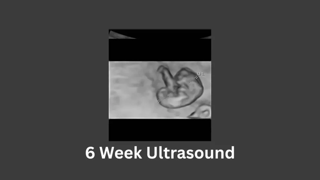
6 Weeks Neural tube closes
By 6 weeks, the development has advanced enough that a transvaginal ultrasound often reveals a tiny embryo (fetal pole) with a heartbeat. A 6-week embryo typically measures around 4–5 mm CRL. On the 6-week ultrasound, you might see a small, flickering heartbeat – though sometimes it’s still a few days too early, and the heartbeat becomes evident closer to 6.5–7 weeks. The embryo’s shape may be visible as a curved tadpole-like structure. Small buds become arms and start to form a c shaped curve.
At this stage, structures like the head and trunk begin to form. If a heartbeat isn’t seen yet, your provider will likely suggest scanning at 7 weeks when the odds of detecting it are much higher. Hearing that first heartbeat is a big milestone of the first trimester. The neural tube of baby are closing.
7 Weeks Head develops
At 7 weeks, an ultrasound usually confirms a viable pregnancy with an embryo and heartbeat. The embryo has grown to about 1.0–1.3 cm (10–13 mm) CRL by now. The embryo often looks like a small bean with a pulsing heartbeat. You may see the outline of the head and the beginnings of limb buds.
The amniotic sac surrounding the baby is expanding and measures only a few millimeters across at this point. This scan reassures that the pregnancy is “viable” (developing normally in the uterus with a heartbeat). In fact, limb development is underway – arm buds are more developed by week 7.
8 Weeks Nose forms
By 8 weeks, the embryo is becoming more recognizable as a tiny baby. 8-week ultrasounds typically show a larger embryo about 1.5 cm (15 mm) long. The head is proportionally large, and the facial features are starting to form, though still not clearly visible. You might observe the little limb buds (early arms and legs) moving – embryos can start to make spontaneous movements around 8 weeks, which can sometimes be seen on ultrasound. The heart is beating strongly, and the sonographer will measure the crown-rump length (CRL) to confirm gestational age.
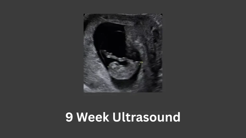
9 Weeks Toes appear
At 9 weeks, the embryo officially transitions to a fetus. A 9-week pregnant ultrasound reveals a growing little one now measuring around 2.0–2.5 cm (about 1 inch) CRL. The baby’s shape is clearer: the head is still large relative to the body, and you can discern the beginnings of ears and nose. The tail nub present in earlier weeks has disappeared by week 9. On ultrasound, the fetus might be seen curling and uncurling or making tiny movements.
10 Weeks The elbows bend
By 10 weeks, the fetus is around 3–3.5 cm long (approximately 1.25 inches) from crown to rump. A 10-week ultrasound shows that the fetus’s head is still proportionally large (brain development is rapid now), and the forehead may appear prominent. The facial features are taking shape – you might see a profile of the tiny nose and lips soon. The body is straightening out from the earlier curled posture. Fingers and toes are forming (though likely too small to visualize clearly yet). At this stage, the ultrasound is often done transabdominally (on your belly) instead of transvaginally, as the fetus and uterus are larger and more easily seen through the abdominal wall.
11 Weeks Genitals develop
At 11 weeks, the fetus is roughly 4–5 cm long. An 11-week ultrasound might zoom in on active little limbs – in fact, the legs and feet are now well-formed. One example image at 11 weeks shows the legs bent at the knees, tiny feet clearly visible. The bones are starting to calcify, so the thigh and shin bones can sometimes be seen as bright lines on the ultrasound.
The nuchal translucency (NT) ultrasound may be scheduled soon (typically done at 11–13 weeks). This is a specialized scan (often part of the 12-week exam) where the sonographer measures the fluid at the back of the baby’s neck as a screening for chromosomal conditions. The placenta is also visible by now as a disk-like structure attached to the uterine wall, and blood flow from the placenta through the umbilical cord can be observed on Doppler modes.
12 Weeks: Fingernails form
12 weeks mark the end of the first trimester. A 12-week ultrasound is often the first routine scan offered if you haven’t had any earlier ones. At 12 weeks, the fetus measures around 5–6 cm (about 2 inches) long. The ultrasound image now clearly shows the baby’s head and torso. You can even see developing facial bones – the skull, jaw, and tiny nose and mouth are visible in profile. You’ll also see the flickering heart and might glimpse the baby moving its arms or sucking its thumb. At this stage, all major organs are formed and the sonographer can check the anatomy in a preliminary way. The placenta’s position is noted (to ensure it’s not covering the cervix) and the number of babies is confirmed (important in case of twins).
(Note: Most routine prenatal care includes an ultrasound around 11–13 weeks for dating and NT screening, and another around 18–20 weeks for detailed anatomy. In a low-risk pregnancy, additional first-trimester ultrasounds beyond 12 weeks may not be needed unless there are concerns.)
Second Trimester (Weeks 13–27)
The second trimester is often when expectant parents get to see more defined images of their baby and perhaps learn the baby’s sex. From week 13 through week 27, the fetus grows from about the size of a peach to the size of a head of cauliflower! Ultrasounds in this period include the standard mid-pregnancy anatomy scan (usually ~20 weeks) and sometimes growth scans or gender determination scans. During the second trimester, different measurements like BPD (biparietal diameter of the head), head circumference, abdominal circumference, and femur length become useful to track growth.
13 Weeks: Bones start to harden
At 13 weeks, the fetus is around 7–8 cm long and weighs about 25 grams. A 13-week ultrasound still resembles the first trimester in many ways, but the fetus is getting bigger and more active. One ultrasound view from above the head at 13 weeks showed the two halves of the developing brain distinctly visible. This highlights that the brain’s basic structure is formed (the cerebrum’s hemispheres). By this week, the baby’s neck is lengthening, so the head is no longer resting on the chest as much. The 13-week mark is also when the fetus’s genitals are forming – skilled ultrasound eyes might guess the sex, but it’s early and not 100% reliable yet. Many parents choose to wait for the mid-trimester scan for confirmation.
14 Weeks Red blood cells form
Week 14 begins the second trimester, and the fetus is about 8–9 cm crown-rump. The arms and legs are longer and start to move in a coordinated fashion. If you are carrying twins, a 14-week ultrasound will clearly show two fetuses and two gestational sacs (if fraternal) or two babies in one sac (if identical). This early second-trimester scan can check that both twins are growing well. By now, the placenta is fully formed and taking over nourishment – you might see the placenta on the scan as a thickened area on the uterine wall. The baby’s heartbeat is strong and typically around 140–160 bpm at this stage.
15 Weeks: Bone development continues
At 15 weeks, the fetus is roughly 10 cm long (4 inches) from crown to rump. The long bones in the legs (femur, tibia) are clearly distinguishable by 15 weeks. An ultrasound image might zoom in on a tiny foot – the toes are separated, and the foot’s outline is cute and obvious. The baby’s skin is still thin, so blood vessels and internal organs can sometimes be faintly seen with advanced ultrasound techniques. You might catch your baby sucking their thumb on the ultrasound, as the sucking reflex is developing.
16 Weeks Eyes move
By 16 weeks, the baby is about 11–12 cm long (head to rump) and weighs around 100 grams. You might see the baby’s umbilical cord on the scan now – a 16-week image can clearly show the cord connecting the baby to placenta, looking like a twisting rope. The cord contains three vessels (which the sonographer will have noted during the anatomy scan later) and carries nutrients and oxygen to your baby. This transvaginal scan checks that the cervix is long and closed. It’s often done between 16 and 20 weeks in high-risk cases.
On the baby’s side, 16 weeks is around when the facial profile becomes well-defined – you can see the nose, lips, and maybe the baby opening and closing its mouth. The baby’s bladder might be seen too (babies start drinking amniotic fluid and urinating by now!). All these details show how active and developed your little one is becoming.
17 Weeks Pregnant Ultrasound
At 17 weeks, the baby is about 12–13 cm CRL (about 5 inches) and weighs nearly 150 grams. At this stage, 3D/4D ultrasound technology can really shine: a 17-week 3D scan can show a clear image of the baby’s face starting to fill out. If the sonographer uses Doppler, you can see the blood flow in color, especially in the umbilical cord or heart chambers. If you haven’t already, you might find out the sex of the baby around this time, as the genitalia are developed and usually distinguishable on screen.
18 Weeks Pregnant Ultrasound
The fetus is about 14 cm (5.5 inches) long, crown-rump, and weighs ~200 grams. At this stage, they can measure the biparietal diameter (BPD) of the head, the head circumference, abdominal circumference, and femur length to ensure growth is on track. Many parents find out if they’re having a boy or a girl – by 18 weeks, a skilled ultrasound tech can usually tell the sex if you want to know (the genital organs are well-formed, and in boys, the penis and scrotum can be seen; in girls, the labia can be visualized).
19 Weeks Pregnant Ultrasound
At 19 weeks, the fetus is around 15 cm long (crown to rump) and weighs ~240 grams. At 19 weeks, an important area to examine is the placenta and its position. In some ultrasound views at 19 weeks, the placenta itself is the focus – appearing as a gray, granular organ attached to the uterine wall. The sonographer checks that the placenta is functioning and not covering the cervix (placenta previa). They also identify the insertion of the umbilical cord into the placenta to ensure it’s normal. The baby’s skin is still thin, but starting to develop a protective coating (vernix).
(Special note: The 20-week anatomy scan is a pivotal ultrasound in the second trimester. It is typically done between 18 and 22 weeks of pregnancy. So whether at 19 weeks, 20 weeks, or 21 weeks, the content of the scan is similar – a comprehensive check of fetal anatomy and growth.)
20 Weeks Pregnant Ultrasound
The fetus is roughly 16 cm (6.3 inches) CRL, or about 25 cm (10 inches) long from head to toe, and weighs around 300 grams. The sonographer will methodically capture images of the baby’s heart, brain, spine, face, stomach, kidneys, limbs, and more. By 20 weeks, most parents who want to know the sex can find out, since the genitals are visible if the baby cooperates. The ultrasound can also estimate fetal weight at this point using formulas (Hadlock formula) based on measurements. If any issues are suspected (like a low-lying placenta or a soft marker on the baby), your provider will discuss follow-ups. Otherwise, after the 20-week scan, many moms won’t have another ultrasound until later in the third trimester.
21 Weeks Pregnant Ultrasound
By 21 weeks, the baby is growing fast – around 18 cm (7 inches) CRL, possibly 27 cm (11 inches) head-to-heel, and weighs ~350 grams. If your anatomy scan is scheduled around now, it will cover the same checks described above. The baby’s skeletal system is more pronounced – you’ll see the curvature of the spine clearly, and ribs may show up as white lines. This week is also around when the uterus and belly are getting big enough that ultrasound visibility remains good, but keep in mind that as the baby grows larger in the third trimester, space gets tight and full-body views won’t be possible.
22 Weeks Pregnant Ultrasound
At 22 weeks, the fetus is about 19 cm CRL (~7.5 inches) and weighs around 430 grams. A 22-week ultrasound continues to show remarkable detail. The ears are fully formed and stand out from the head. So at 22–23 weeks, you might catch similar behavior on the scan. By 22 weeks, the fetal weight can be estimated to be close to a pound, and the baby’s body is getting more proportional (the legs and arms have lengthened considerably since the first trimester).
23 Weeks Pregnant Ultrasound
At 23 weeks, the baby is about 20 cm CRL (~8 inches) and around 500+ grams (over 1 pound). A 23-week ultrasound is often not routine unless there’s a specific reason (many anatomy scans would have been done by now). The skin of the baby is still translucent, but fat is beginning to accumulate. Meanwhile, the baby’s hearing is improving; loud sounds might make the baby jump, which you could actually see on an ultrasound if it coincided with a noise. Medically speaking, 23 weeks is just below the generally accepted viability threshold.
24 Weeks Pregnant Ultrasound
The fetus at 24 weeks is about 21 cm CRL (~8.5 inches) or ~30 cm (12 inches) head-to-heel, and weighs around 600–630 grams. A 24-week pregnant ultrasound will show a fairly active baby with well-defined bones. By this stage, the baby’s fingerprints have formed (though you can’t see those on ultrasound, of course!). Babies can also respond to touch; during an ultrasound, they might kick or move if they feel the probe pressing on the belly. Internally, the lungs are developing but are not mature.
25 Weeks Pregnant Ultrasound
At 25 weeks, the fetus is about 22 cm (8.8 inches) CRL and ~34 cm (13.5 inches) head-to-heel, weighing around 700 grams. A 25-week ultrasound might be performed in certain situations (like if you had an anterior placenta and they want to check it, or if the baby’s position obscured some anatomy at the 20-week scan and a follow-up is needed). The spine is very prominent on ultrasound now – one image at 25 weeks highlighted the baby’s spine curving along the length of the torso. The vertebrae show up as bright, repeating segments. This is also a good time to check the umbilical cord vessels with Doppler if needed, or to observe the placental position again.
26 Weeks Pregnant Ultrasound
At 26 weeks, the baby is about 23 cm (9 inches) CRL and ~35 cm (14 inches) long head-to-foot, weighing roughly 800 grams. By 26 weeks, the baby’s lungs are practicing breathing – they inhale and exhale amniotic fluid. On an ultrasound, you might actually see the baby’s chest make breathing movements. In a described ultrasound at 26 weeks, the baby opened his mouth wide – possibly swallowing fluid or yawning. The retinas of the eyes are forming; by next week, the eyes will open. The brainwave activity is maturing – babies at this stage have sleep and wake cycles. Unless there’s a medical indication, not everyone gets an ultrasound at 26 weeks.
27 Weeks Pregnant Ultrasound
The fetus is about 24 cm (9.5 inches) CRL, or around 36–37 cm (14.5 inches) from head to toe, and weighs about 900–1000 grams (nearly 2.0 lbs). By 27 weeks, an ultrasound can clearly show fine details like the blood flow in certain vessels, especially using Doppler. This week on ultrasound, you might also see the baby’s eyes opening and closing; yes, around 27–28 weeks, babies start opening their eyes, although there’s not much to see in the dim uterus! Doctors may measure the amniotic fluid index (AFI) if needed to ensure levels are normal.
Third Trimester (Weeks 28–40+)
Welcome to the third trimester! From week 28 through delivery (which is usually around 40 weeks, though it can be a bit earlier or later), ultrasounds are typically done on an as-needed basis. Common reasons include checking fetal growth, placenta position, fluid levels, or baby’s position (head-down or breech). In some pregnancies, a 28-32 week growth scan is done, and many providers also do a scan at around 36 weeks to ensure the baby is positioned head-down. Additionally, ultrasounds can be part of biophysical profiles (BPP) in the late third trimester to assess fetal well-being (especially if you go overdue or have risk factors).
28 Weeks Pregnant Ultrasound
At 28 weeks, you are officially in the third trimester. Baby is about 25 cm (10 inches) CRL, roughly 38 cm (15 inches) head-to-foot, and weighs around 1.0–1.1 kg (about 2.5 lbs). This is also an optimal time for elective 3D/4D ultrasounds because the baby has some fat but still enough room to be seen well. On an ultrasound at 28 weeks, you can see both blood flow and fluids in action. The baby’s kidneys and bladder should be visible and functioning. The heart rate is usually around 130–140 bpm now and easy to hear. If your baby is head-down already, you might see the round contour of the head low in the uterus; if the baby is breech (head up), the head will appear at the top. Doctors will note the baby’s position at this scan but it may change in the coming weeks.
29 Weeks Pregnant Ultrasound
By 29 weeks, the baby is around 26 cm (10.5 inches) CRL and ~39-40 cm (16 inches) long overall, with a weight of approximately 1.2–1.3 kg (about 2.7–3 lbs). For instance, you might see a little foot pressed up and perhaps kicking against the uterine wall. It’s common for moms to start feeling stronger kicks in the ribs around this time, and indeed, an ultrasound may show a foot up near the ribs. The bones are fully developed (though still soft), so those kicks have power! They might mention percentiles (for example, the baby might be at the ~50th percentile weight for 29 weeks).
30 Weeks Pregnant Ultrasound
At 30 weeks, baby is approximately 27 cm (10.8 inches) CRL, around 40 cm (16 inches) in total length, and weighs roughly 1.3–1.4 kg (about 3 lbs). The position of the baby is an important thing around 30 weeks: many babies will start to settle head-down (vertex) by this time, though some still flip. The ultrasound will note if baby is head-down, breech, or transverse. The amniotic fluid volume usually peaks around 32 weeks, so at 30 weeks, there’s still a decent amount of fluid around the baby – you might see large pockets of fluid on the scan, which is normal.
31 Weeks Pregnant Ultrasound
At 31 weeks, baby is about 28 cm (11 inches) CRL, ~41 cm (16.5 inches) long, and weighs around 1.5 kg (3.3 lbs). On a 31-week scan, you can see the baby’s eyes blink and even catch the glint of the lens within the eye when open. If you shine a flashlight on your belly, the baby might turn their head – occasionally, during an ultrasound, if a bright Doppler or probe lights up, the baby might respond. But most babies will be tracking along fine. The lungs are maturing; if the sonographer does a BPP, they’ll watch for fetal breathing movements (seeing the baby’s diaphragm “practice breathing”).
32 Weeks Pregnant Ultrasound
At 32 weeks, the baby is around 29 cm (11.5 inches) CRL, about 42–43 cm (17 inches) tall, and weighs approximately 1.7–1.8 kg (3.7–4.0 lbs). Hair might be visible on the head; in fact, at 25-26 weeks, some ultrasound techs can see hair floating in the amniotic fluid if the baby has a lot (it appears as fuzzy white strands on a detailed scan). Doctors start paying attention to the position of the baby now. If fluid is too high or too low, interventions may be considered. Also, if the baby was breech earlier, they might have flipped by 32 weeks – about 90% of babies are head-down by 32 weeks, though some take a bit longer.
33 Weeks Pregnant Ultrasound
At 33 weeks, the baby is about 30 cm (12 inches) CRL, ~44 cm (17.5 inches) in total length, and weighs around 1.9–2.0 kg (about 4.2 lbs). The kick counts are important now (you should be monitoring the baby’s movements daily). During a BPP at 33 weeks, the tech would look for things like movements, tone (baby flexing arms/legs), practice breathing, and adequate fluid. Usually, babies pass these tests just fine. By 33 weeks, the immunity from mom is being passed to the baby, and the baby’s lungs are pretty well developed (though not fully mature). The umbilical cord blood flow might be measured via Doppler if there’s any growth concern.
34 Weeks Pregnant Ultrasound
At 34 weeks, the baby is about 31 cm (12.2 inches) CRL, ~45 cm (18 inches) long head-to-foot, and weighs roughly 2.2 kg (4.8 lbs). On a 34-week ultrasound, one cute detail that can be seen is the baby’s ear. For instance, an image might zoom in on a fully formed little ear. The cartilage in the ear is firmer now, and the ear is in the right place on the head (ear positioning can be a marker they check in some syndromes, but by 34 weeks, everything looks normal and developed). Everything is getting set for the last few weeks of pregnancy.
35 Weeks Pregnant Ultrasound
At 35 weeks, the baby is around 32 cm (12.5 inches) CRL, ~46 cm (18+ inches) long, and weighs about 2.5 kg (5.5 lbs). But if not fully engaged, you might still catch glimpses of that sweet face pressing against the uterine wall. At 35 weeks, doctors are interested in the cervix if preterm labor signs exist – sometimes a cervical length check is done if there are contractions. Otherwise, ultrasound might be used to estimate fetal weight, especially if any plan is needed (like deciding on inducing early if the baby is measuring very large or if there’s growth restriction). A BPP or non-stress test might be ordered weekly from 36 weeks on if you have conditions like gestational diabetes or high blood pressure, and an ultrasound plays a role in those assessments.
36 Weeks Pregnant Ultrasound
36 weeks is often when the final preparations for birth begin. Baby is about 33 cm (13 inches) CRL, ~47 cm (18.5 inches) long overall, and weighs roughly 2.7–2.8 kg (around 6 lbs). Many providers will do a quick 36-week ultrasound to verify baby’s position (since by 36 weeks the baby should ideally be head-down for a vaginal delivery). One ultrasound image at 36 weeks showed a baby girl’s genital area, confirming gender (by now, that’s very clear). The amniotic fluid index is often measured at 36 weeks to ensure you don’t have low fluid (oligohydramnios) or too much (polyhydramnios).
37 Weeks Pregnant Ultrasound
At 37 weeks, your pregnancy is considered “full-term” (early term, technically). Baby is around 34 cm (13.5 inches) CRL, ~48–49 cm (19 inches) tall, and weighs roughly 3.0 kg (6.6 lbs) – though there is wide variation. Generally, there is no routine ultrasound at 37 weeks unless an issue is suspected. However, if you go for a 37-week ultrasound, it might be part of a biophysical profile (BPP) or just a final check on the baby’s well-being. The baby’s position at 37 weeks should definitely be head-down in most cases. If the baby is breech at 37 weeks, a decision for version or C-section is imminent. On the ultrasound, the baby’s body looks quite cramped – you’ll see that there’s not a lot of space, and the baby’s knees and elbows are often bent in close. If there are concerns about movement, a non-stress test and BPP are common; the ultrasound portion will ensure the baby’s breathing movements and tone are good. At 37+ weeks, if everything is normal, you might not see the baby via ultrasound again – next time will be in person!
38 Weeks Pregnant Ultrasound
At 38 weeks, the baby is around 35 cm (14 inches) CRL, ~50 cm (20 inches) in length, and weighs about 3.2–3.3 kg (7 lbs). Most pregnancies won’t require an ultrasound at 38 weeks unless you have a specific indication (like hypertension in pregnancy, where they might do weekly BPPs). Usually, the baby will pass easily – for instance, the sonographer might watch for 30 minutes to see the baby practice breathing or move. If the baby is a bit “sleepy,” sometimes drinking something cold or sugary can reduce motion. But for most, ultrasound simply confirms all is well.
39 Weeks Pregnant Ultrasound
At 39 weeks, the baby is about 36 cm (14.5 inches) CRL, ~51 cm (20+ inches) long, and weighs roughly 3.4–3.5 kg (7.5 lbs). Ideally, you’re in the final days of pregnancy. There is typically no ultrasound at 39 weeks in an uncomplicated pregnancy – your provider is probably checking your cervix and monitoring fetal heart rate with a Doppler instead. Ultrasound might also estimate weight if there’s concern about macrosomia (very large baby) – though at this stage, weight estimates can be a bit off. If the baby is estimated, say, 9+ pounds at 39 weeks, your provider might discuss delivery options, but many times spontaneous labor is still awaited.
One more important check at 39 weeks is the baby’s position relative to the birth canal: ultrasound isn’t typically used for this (manual exam is), but if the baby’s head seems large or not well positioned, some might measure head circumference or do a quick scan to reassure everything looks favorable. At this point, everyone is simply anticipating the safe arrival of your baby!
40 Weeks Pregnant Ultrasound
40 weeks is your due date (though only a minority of babies arrive exactly this week!). Baby is around 37 cm (14.5–15 inches) CRL, ~51–52 cm long (~21 inches), and weighs roughly 3.5–3.6 kg (about 7.7–8 lbs) on average. A 40-week ultrasound often focuses on amniotic fluid levels and placental condition. Because once you reach 40 weeks and beyond, the placenta can start to age and the fluid can decrease, your provider will want to ensure the baby is still well-nourished and safe. If the baby has dropped into the pelvis, the head might be so low that ultrasound visualization is limited. If any signs of placental insufficiency or distress show (like low fluid or poor movements), many doctors will recommend delivering (since the baby is fully term).
41–42 Weeks (Post-term) Ultrasound
If your pregnancy extends beyond 40 weeks, you enter the post-term period (41 and 42 weeks). At 41 weeks, and certainly by 42 weeks, you will likely have close monitoring, which can include an ultrasound. A 41-week ultrasound typically checks that the baby is still doing well in utero, measuring fluid levels and doing a BPP. Many providers induce labor by 41 weeks or at least by 42, as the placenta’s function can decline past 42 weeks. At this stage, if the baby has not born, an ultrasound might estimate the baby’s weight one more time and confirm the baby’s head is well down. Sometimes a 41-week ultrasound can catch the baby’s position, such as if the head is molded or engaged in the pelvis. Rest assured that by 41-42 weeks, the baby’s development is complete; they’re mostly just gaining a bit more weight (sometimes a few extra ounces) and perhaps growing longer hair and nails!
Throughout this week-by-week journey, ultrasounds serve as invaluable tools – from confirming a tiny heartbeat in the first trimester to monitoring growth and well-being in the final weeks. These scans not only provide medical insights but also allow expectant parents to bond with their baby by seeing those first images in the womb. Each milestone, whether a 7-week heartbeat flicker or a 20-week anatomy revelation or a 36-week position check, marks progress in the pregnancy.
In conclusion, pregnancy ultrasounds by week show the amazing progression from a microscopic embryo to a fully formed baby. These images and measurements guide obstetrical care, helping confirm due dates, track development, and make critical decisions when needed. Coupled with tools like the conception and due date calculator, they equip you with knowledge about your pregnancy timeline. With each passing week, the ultrasound transforms from a faint speck on the screen to the unforgettable sight of your baby’s face – reinforcing the reality of new life and the short countdown until you finally meet your little one in person.