Immunology Made Simple

1. Spleen

1. Location and Immune Response

  • LUQ (Left Upper Quadrant):
    • Location of the spleen.
  • Beta and T Cells:
    • Beta cells: Located in the pancreas (in the islets of Langerhans) and involved in insulin production.
    • T cells: Part of the immune system, involved in adaptive immunity.

2. Infectious Mononucleosis:

  • Cause:
    • Epstein-Barr Virus (EBV).
  • Clinical Features:
    • Splenomegaly: Enlargement of the spleen due to infection.
    • T-cell Hyperplasia: An increase in T cells as part of the immune response to the viral infection.
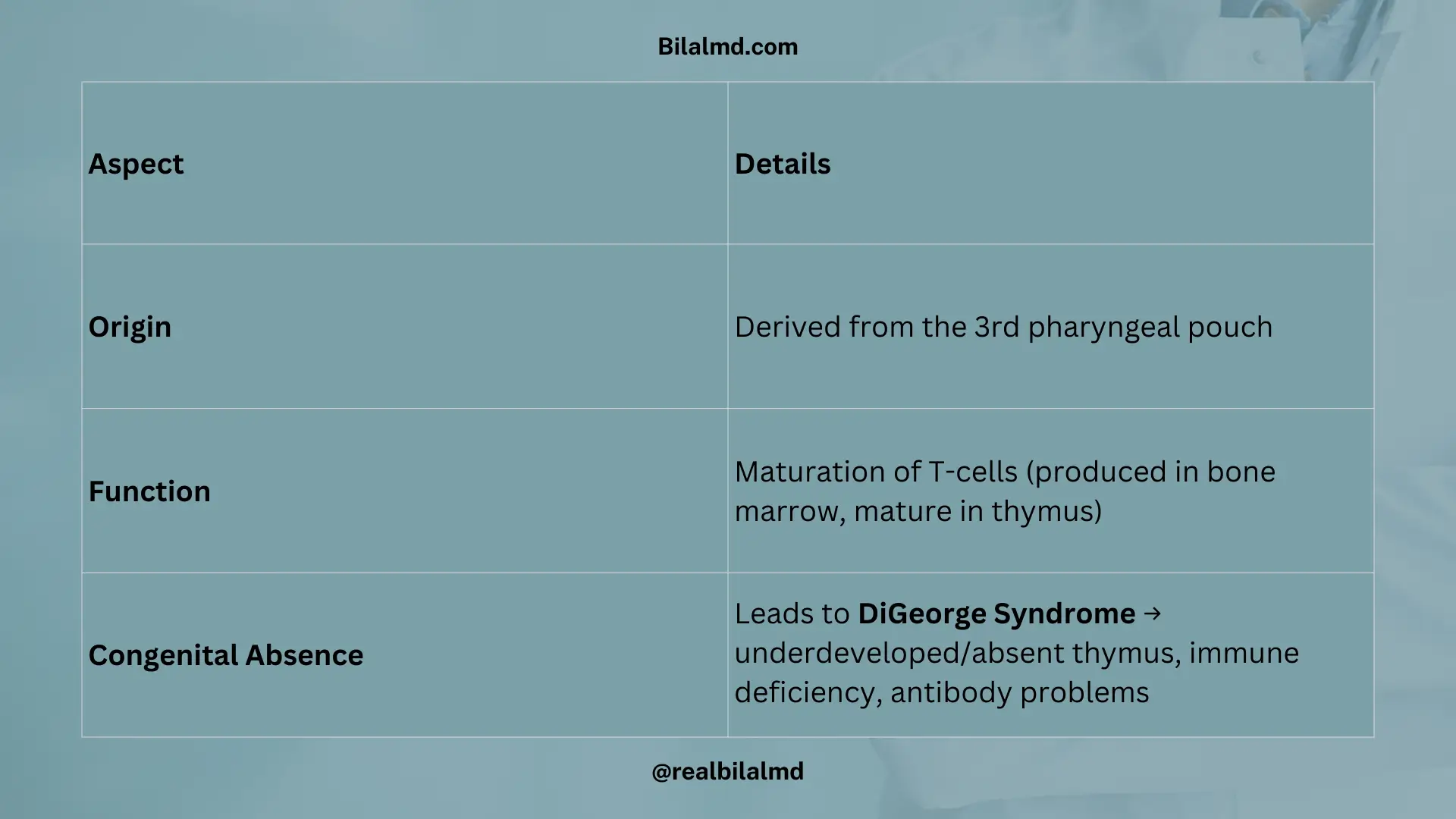
Spleen location, immune role, and splenomegaly in EBV infection

2. Thymus

  1. Origin:
    • Derived from the 3rd pharyngeal pouch.
  2. Function:
    • Responsible for the maturation of T-cells.
    • T-cells are made in the bone marrow, but they mature in the thymus.
  3. Congenital Absence:
    • If the 3rd pharyngeal pouch is absent, it leads to DiGeorge Syndrome, a condition where the thymus is underdeveloped or absent, affecting immune function and causing antibody deficiencies.
Thymus embryology, T-cell maturation, and DiGeorge syndrome

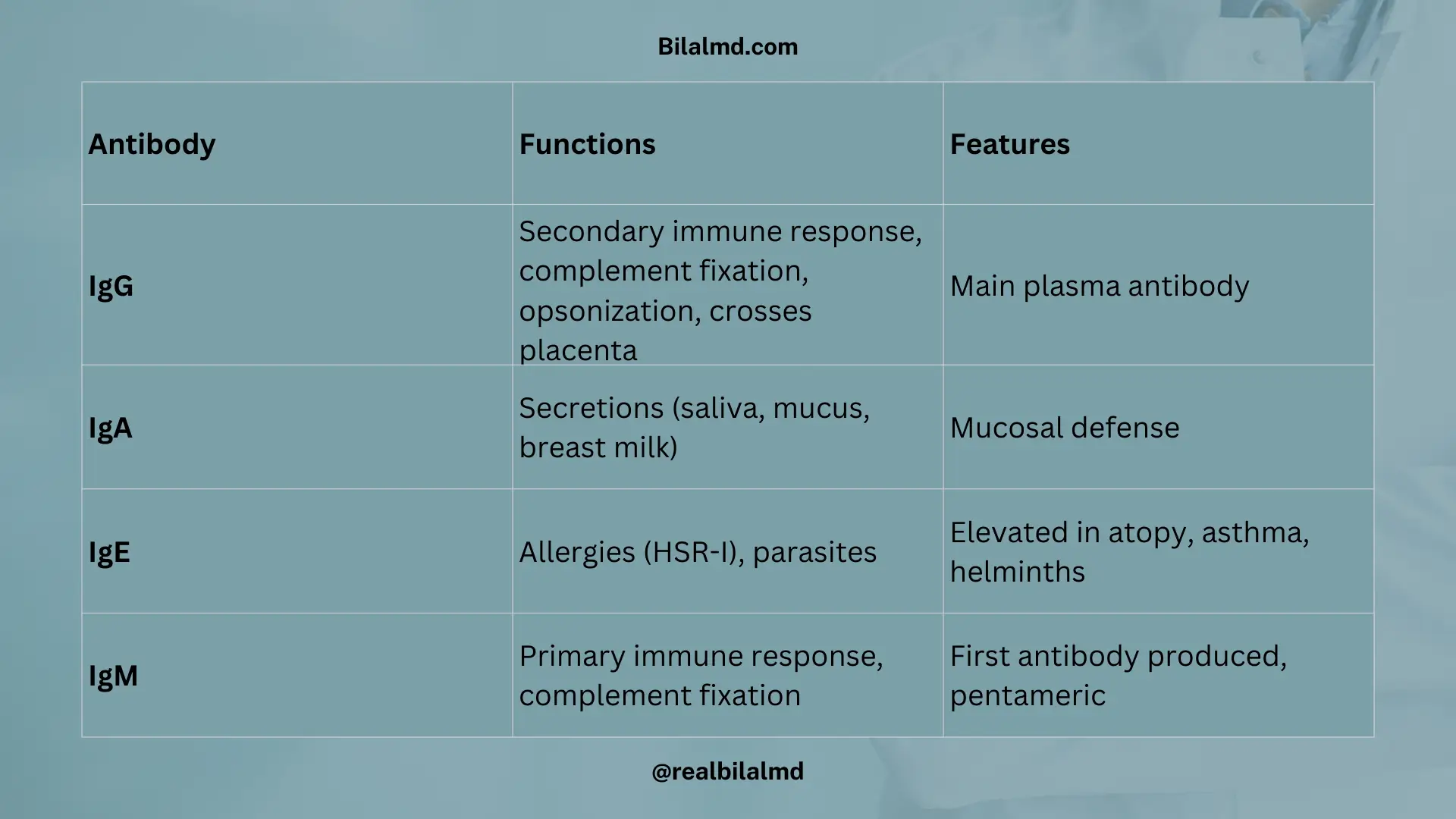
3. Antibodies and Immune Responses

AntibodyFunctionCharacteristics
IgGSecondary immune responseCrosses the placentaComplement fixationOpsonizationMore in plasma– Main antibody in secondary immune response – Helps fix complement and aids in opsonizationCrosses placenta to provide passive immunity
IgA– Present in secretions (saliva, mucus, breast milk) – More produced but drains– Found in mucosal membranes – Lower concentration in blood but crucial for mucosal immunity
IgE– Involved in HSR-1 (Type I hypersensitivity) – Plays a role in parasitic infections– Involved in allergic reactions and parasite defenseElevated in parasitic infections and allergies
IgMPrimary immune responseFixes complementPentameric structureFirst antibody produced during primary responseFixes complement and activates immune response
IgG, IgA, IgE, and IgM functions in immunity

4. Types of Grafts

Graft TypeDefinitionExample
AutograftGraft taken from the same bodySkin graft from one part of the body to another part of the same body
IsograftGraft between identical twins (genetically identical)Organ transplant between identical twins
AllograftGraft between individuals of the same speciesKidney transplant between non-identical humans
XenograftGraft between individuals of different speciesHeart valve transplant from a pig to a human
Types of grafts in transplantation with examples

5. Transplant Rejection

Type of RejectionTimeframeDescription
Acute RejectionOccurs within weeks to months– The immune system attacks the transplanted organ shortly after the transplant.
Chronic RejectionOccurs within months to years– Long-term rejection that leads to gradual deterioration of the transplanted organ over time.
Hyperacute RejectionOccurs within minutes– Immediate rejection, often due to pre-existing antibodies against the donor tissue, resulting in rapid organ failure.
Hyperacute, acute, and chronic transplant rejection differences

6. HSR-I (Type I Hypersensitivity Reaction)

Type of HypersensitivityMechanismDetails
HSR-I (Type I)IgE-mediatedGenetic predisposition (atopy). – Mast cells release histamine upon activation by IgE. – Conditions: Severe anaphylaxis, asthma, atopic dermatitis, allergic rhinitis.
HSR-II (Type II)Antigen + Antibody Complex → Cell damageIgG or IgM antibodies bind to antigens on the cell surface, leading to cell damage (e.g., hemolytic anemia, autoimmune diseases).
HSR-III (Type III)Antigen + Antibody + Complement Activation– Formation of immune complexes, leading to complement activation and tissue damage (e.g., in lupus, rheumatoid arthritis).
HSR-IV (Type IV)T-cell MediatedT-cell mediated immune response causes tissue damage (e.g., in contact dermatitis, tuberculosis, graft rejection).
Type I, II, III, IV hypersensitivity reactions with examples

7. Complement System

  • Helps to reduce infection.
  • Group of 20 proteins.
  • Proteins are inactive, become active during infection.
  • 11 proteins are important.
  • Each complement protein has 2 components.
    • Example: C1 → C1a, C1b.
  • Linked with T-cell mediated hypersensitivity type IV.

1. Pathways of Activation

  • Classical pathway → triggered by IgG or IgM.
  • Alternative pathway → triggered directly by microbial surfaces.
  • Mannose-binding lectin pathway → triggered by mannose residues on microbes.

2. Important Functions

  • Opsonization → C3b coats microbes, enhances phagocytosis.
  • Chemotaxis → C5a attracts neutrophils.
  • Membrane Attack Complex (MAC) → C5b, C6, C7, C8, C9 assemble and lyse microbes.

3. Deficiency States

  • C5–C9 deficiency → recurrent Neisseria bacteremia.
  • C1–C6 deficiency → recurrent pyogenic infections (pus-forming bacteria), ↑ risk of SLE.

4. Adaptive Immunity Link

  • B-cell maturation → Plasma cells → Antibody production → Humoral immunity.
  • T-cells → Cell-mediated immunity.
    • CD4 (Helper T cells) → Support humoral immunity.
    • CD8 (Cytotoxic T cells) → Directly kill infected cells.
Complement activation pathways, functions, and deficiencies

Here are other materials for NLE NRE step 1

5. Innate Immunity

  • No memory, acts immediately and non-specifically.
  • Components:
    • Physical barriers (skin, mucosa).
    • Cells: Neutrophils, Macrophages, NK cells.
    • Proteins: Complement system, acute phase proteins.

6. Adaptive Immunity

  • Specific response develops memory.
  • Components:
    • B-cells → mature into plasma cells → produce antibodies.
    • T-cells → CD4 (helper) and CD8 (cytotoxic).
  • Basis of therapeutic monoclonal antibodies.
Differences between innate and adaptive immunity

1. Monoclonal Antibodies (Examples)

DrugTargetUse
DenosumabRANK-LOsteoporosis
OmalizumabIgEAsthma (allergic)
PalivizumabRSV F proteinRSV bronchiolitis prophylaxis
EculizumabC5 complementParoxysmal nocturnal hemoglobinuria (PNH)
EfalizumabCD11a (LFA-1)Psoriasis
Natalizumabα4-integrinMultiple sclerosis
TrastuzumabHER2/neu receptorHER2+ breast cancer, gastric cancer

Check your NRE Step 1 result after completing the exam.

Similar Posts

  • Reproductive Cancer Made Simple

    1. Prostate Cancer Here are other materials for NLE NRE step 1 2. Testicular Cancer 3. Germ Cell Tumors Tumor Type Tumor Marker Yolk Sac Tumor AFP (Alpha-fetoprotein) Choriocarcinoma Beta-HCG Teratoma AFP or Beta-HCG Seminoma Negative for AFP and Beta-HCG 4. Non-Germ Cell Tumors Tumor Tumor Marker Leydig Cell Cancer Increased Testosterone Sertoli Cell Cancer…

  • Musculoskeletal Made Simple

    1. Gout 2. Pseudogout Feature Gout Pseudogout Crystal type Monosodium urate (MSU) Calcium pyrophosphate (CPPD) Crystal shape Needle-shaped Rhomboid-shaped Birefringence Negative birefringence Positive birefringence Common site 1st MTP joint (Podagra) Knee joint (most common) Subcutaneous deposit Tophi Rare Serum finding ↑ Uric acid (>7.5 mg/dL) Normal uric acid Acute treatment NSAIDs NSAIDs, Colchicine Chronic treatment…

  • Neonatology Made Simple

    1. Apgar Score A rapid scoring system used to evaluate a newborn’s cardiopulmonary adaptation after birth. Apgar Parameters and Scoring Sign 0 Points 1 Point 2 Points Activity (Muscle tone) No movement Arms flexed, legs extended Active movement Pulse (Heart rate) Absent <100 beats/min >100 beats/min Grimace (Reflex irritability) No response Some flexion of extremities…

  • Surgery Made Simple

    1. Burn Tissue Injury 1. Types 2. Common Infection 3. Fluid Resuscitation Parkland Formula Fluid (ml)=4[Weight (kg)×%BSA(burned)] 🔹 Example:Weight = 70 kg, Burn area = 5% BSA 4×70×5=1400 ml Here are other materials for NLE NRE step 1 2. Shock Mnemonic for shock types → SHOCk 1. Hypovolemic Shock 2. Obstructive Shock 3. Cardiogenic Shock 4. Distributive Shock…

  • GIT Made Simple

    Digestive System (GIT) – Medicine Esophagus: Length: 25 cm long. Strongest Layer: Submucosa. Immunocompromised + Dysphagia + Endoscopy: White patches on endoscopy indicate fungal infection, likely fungal esophagitis. Symptoms: Barium Imaging: Diagnosis: Esophageal Cancer: Barrett’s Esophagus: Region Arteries Cervical Inferior thyroid artery, subclavian artery Thoracic Descending aorta Abdominal Left gastric artery 2. Stomach Shape: J-shaped…

  • Renal Made Simple

    1. Nephrotic vs Nephritic Syndrome Nephrotic Syndrome Causes: Nephritic Syndrome (Inflammatory): Post-infectious: 2. Vasculitis ANCA-associated: Anti-GBM: Hereditary nephritis: 3. Acid–Base Balance & ABGs 4. Polycystic Kidney Disease (PKD) Feature ARPKD ADPKD Onset Childhood Adult Frequency Rare Common Prognosis Poor Good Association Liver cysts Berry aneurysm (↑ SAH risk) USG Enlarged kidneys w/ cysts Multiple cysts…

Leave a Reply

Your email address will not be published. Required fields are marked *